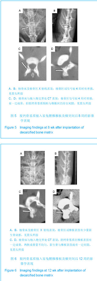
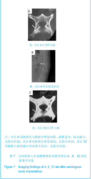
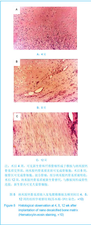
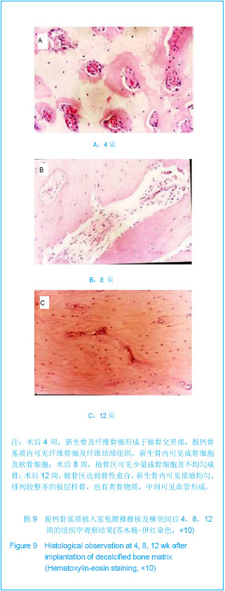
| [1] Dinopoulos H,Dimitriou R, Giannoudis PV. Bone graft substitutes: What are the options? Surgeon.2012; 10(4): 230-239.[2] Bhatt RA,Rozental TD.Bone Graft Substitutes.Hand Clin. 2012;28(4): 457-468.[3] Enrique Guerado, Carl Hans Fuerstenberg. What bone graft substitutes should we use in post-traumatic spinal fusion? Injury.2011;42 Suppl 2: S64-S71.[4] Seebach C,Schultheiss J,Wilhelm K,et al.Comparison of six bone-graft substitutes regarding to cell seeding efficiency, metabolism and growth behaviour of human mesenchymal stem cells (MSC) in vitro. Injury.2010;41(7): 731-738.[5] Nenad I,Petar N,Zorica A,et al. Biphasic calcium phosphate coated with poly d,1 lactide co glycolide hiomaterial as a hone substitute. Eur Ceram Soc.2007;27: 1589-1594.[6] De Long WG Jr,Einhorn TA,Koval K,et al. Bone grafts and bone graft substitutes in orthopaedic trauma surgery.A critical analysis .J Bone Joint Surg Am. 2007;89(3):649-658.[7] Hsu W,Fuchs D.A Meta-Analysis of Fusion Rates from Bone Graft Substitutes in a Rodent Posterolateral Spine Arthrodesis Model.Spine J.2011;11(10 Suppl): S59-S60.[8] Athanasiou VT,Papachristou DJ,Panagopoulos A,et al.Histological comparison of autograft, allograft- DBM, xenograft,and synthetic grafts in a trabecular bone defect:an experimental study in rabbits.Med Sci Monit.2010;16(1): 24-31.[9] Powell M,Griffin M,Tai S. Bottom-up risk regulation? How nano-technology risk knowledge gaps challenge federal and state environmental agencies. Environ Manage.2008;42(3): 426-443.[10] Hak DJ.The use of osteoconductive bone graft substitutes in orthopaedic trauma.J Am Acad Orthop Surg.2007;15(9): 525-536.[11] Janicki P,Schmidmaier G.What should be the characteristics of the ideal bone graft substitute? Combining scaffolds with growth factors and/or stem cells. Injury.2011;42 Suppl 2: S77-S81.[12] Blokhuis TJ,Arts JJ.Bioactive and osteoinductive bone graft substitutes: Definitions, facts and myths.Injury.2011;42 Suppl 2: S26-S29.[13] Jurczyk MU,Jurczyk K,Miklaszewski A,et al.Nanostructured titanium-45S5 Bioglass scaffold composites for medical applications.Mater Design. 2011;32(10):4882-4889.[14] Caldorera-Moore M,Peppas NA.Micro- and nanotechnologies for intelligent and responsive biomaterial-based medical systems.Adv Drug Del Rev.2009;61(15):1391-1401.[15] Rodgers WB, Gerber EJ. A DBM, BMA, Local Bone Graft Composite in Multi-Level PLIF: Fusion Rates. Spine J. 2010; 10(9 Suppl): S26.[16] Li XQ,Liu TQ,Song KD.Dalian Ligong Daxue Xuebao. 2005; 45(5):653-657.李香琴,刘天庆, 宋克东.成骨细胞在纳米材料表面上粘附特性[J].大连理工大学学报,2005,45(5):653-657.[17] Qian Y,Zhang ZF,Shen ZL,et al.Zhongguo Jiaoxing Waike Zazhi. 2005;13(12):911-914.钱鋆,张兆锋,沈尊理,等.冻干脱钙骨表面纳米结构对细胞行为的影响[J].中国矫形外科杂志,2005,13(12):911-914.[18] Yoshikawa H,Myoui A.Bone tissue engineering with porous hydroxyapatite ceramics.Artif Organs.2005;8(3): 131-136.[19] Bohner M.Resorbable biomaterials as bone graft substitutes. Mater Today.2010;13(1-2): 24-30.[20] Zimmermann G,Moghaddam A.Allograft bone matrix versus synthetic bone graft substitutes. Injury.2011;42 Suppl 2: S16-S21.[21] Geurts J,Chris Arts JJ,Walenkamp GH.Bone graft substitutes in active or suspected infection. Contra-indicated or not? Injury.2011;42 Suppl 2:S82-S86.[22] Drosos GI,Babourda E,Magnissalis EA,et al. Mechanical characterization of bone graft substitute ceramic cements. Injury.2012;43(3): 266-271.[23] Chitsazi MT,Shirmohammadi A,Faramarzie M,et al.A clinical comparison of nano-crystalline hydroxyapatite (Ostim) and autogenous bone graft in the treatment of periodontal intrabony defects. Med Oral Patol Oral Cir Bucal. 2011;16(3): E448-E453.[24] Kweon H, Lee KG, Chae CH,etal.Development of Nano-Hydroxyapatite Graft With Silk Fibroin Scaffold as a New Bone Substitute. J Oral Maxillofac Surg.2011; 69(6): 1578-1586.[25] Chai YC,Kerckhofs G,Roberts SJ,et al. Ectopic bone formation by 3D porous calcium phosphate-Ti6Al4V hybrids produced by perfusion electrodeposition. Biomaterials.2012; 33(16):4044-4058.[26] Huang K,Chen XS,Jia LS,et al.Zhongguo Jiaoxing Waike Zazhi. 2009;17(13):1017-1019.黄凯,陈雄生,贾连顺,等. 纳米脱钙骨基质的制备及其性能检测[J].中国矫形外科杂志,2009,17(13):1017-1019.[27] Zhao XM.Dianda Ligong. 2005;27(4):35-36.赵晓明.纳米材料的特性及应用[J].电大理工,2005,27(4):35-36.[28] Qian Y,Shen ZL,Zhang ZF,et al.Zhongguo Xiufu Chongjian Waike Zazhi. 2006;20(5):561-564.钱鋆,沈尊理,张兆锋,等.运用组织工程原理结合纳米技术构建骨组织的实验研究[J].中国修复重建外科杂志,2006,20(5): 561-564.[29] Kailasanathan C,Selvakumar N,Naidu V. Structure and properties of titania reinforced nano-hydroxyapatite/gelatin bio-composites for bone graft materials. Ceran Int.2012;38(1): 571-579.[30] Tang ZB,Cao JK,Wen N,et al.Posterolateral spinal fusion with nano-hydroxyapatite-collagen/PLA composite and autologous adipose-derived mesenchymal stem cells in a rabbit model.J Tissue Eng Regen Med. 2012;6(4):325-336. |