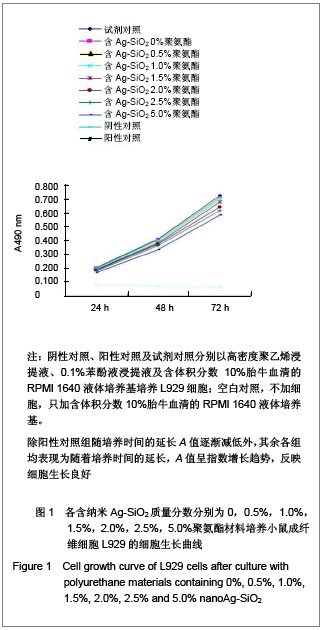
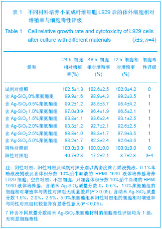
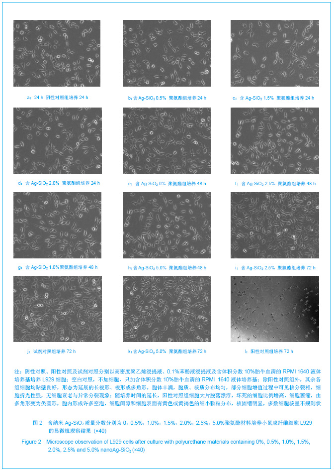
| [1] Zhao HS,Cai HB,Pan ZQ,et al.Gongneng Gaofenzi Xuebao. 2005,18(3):361-367. 赵宏生,蔡海波,潘肇琦,等. 生物载体用聚醚型聚氨酯多孔材料的研究[J].功能高分子学报,2005,18(3):361-367.[2] Wang XM.Reguxing Shuzhi. 2009;24(4):47-49. 王学敏. 医用聚氨酯材料的研究进展及发展方向[J].热固性树脂,2009,24(4):47-49.[3] Tan HM,Wang JX,Zhang JL.Huaxue Tuijinji yu Gaofenzi Cailiao. 2009;7(2):23-26. 谭红梅,汪建新,张俊良.医用聚氨酯的改性及应用[J].化学推进剂与高分子材料,2009,7(2):23-26.[4] Wang WZ,Zhang C,Chen JH.Youjigui Cailiao. 2001;15(5): 33-37. 王文忠,张晨,陈剑华.有机硅改性聚氨酯的研究进展[J].有机硅材料,2001,15(5):33-37. [5] Zhang CM.Shijie Xiangjiao Gongye.2003;31(5):45-48. 张承焱.漫谈医用聚氨酯[J].世界橡胶工业,2003,31(5):45-48.[6] Sang JG,Lu KW,Wang JH,et al.Zhongguo Jiaoxing Waike Zazhi. 2001;8(6):604-605. 桑井贵,鲁凯伍,王建华,等. 医用聚氨酯绷带的临床应用[J].中国矫形外科杂志,2001,8(6):604-605.[7] Li J,Gong ZC,Deng LD,et al.Huaxue Gongye yu Gongcheng. 2001;21(4):235-238. 李军,龚志超,邓联东,等. 皮肤用亲水性聚氨酯压敏胶的制备及性能研究[J].化学工业与工程,2001,21(4):235-238.[8] Liu L,Wei ZY,Gao J,et al. Zhongguo Zuzhi Gongcheng Yanjiu yu Linchuang Kangfu. 2008;12(14):2735-2738. 刘炼,魏志勇,高军,等. 生物可降解聚氨酯的合成及应用[J].中国组织工程研究与临床康复,2008,12(14):2735-2738.[9] Huang ZB,Li BG,Hu Y,et al.Hangtian Yixue yu Yixue Gongcheng. 2001;14(5):355-359. 黄忠兵,李伯刚,胡英,等. 新型亲水性聚氨酯敷料表面界面性能的研究[J].航天医学与医学工程,2001,14(5):355-359.[10] Cheng LP,Hu Y,Zheng CQ,et al.Shengwu Yixue Gongcheng Yanjiu. 2004;23(4):40-43. 程莉萍,胡英,郑昌琼,等. 聚氨酯抗菌创伤敷料的制备及其灭菌效果的研究[J]. 生物医学工程研究,2004,23(4):40-43.[11] Fu W,Wu WH,Wang QP. New Silver-Based Antibacterial Agent Advancement and Progress.Jiangsu Ceram.2006; 39(5):26-28.[12] GB/T 16886.12-2005.[13] Shi GH,Deng C.Mandible bone repair in rabbits with chitosan/n-HA composites. Chin J Modern Med.2007; 17(19):2322-2326.[14] Zou Y,Wang WM,Zhu YN,et al.Kouqiang Yixue Yanjiu. 2011;27(8):673-675. 邹颖,王文梅,朱雅男,等. CCK-8法评价口腔粘结材料的体外细胞毒性研究[J].口腔医学研究,2011,27(8):673-675.[15] Oberdörster G,Sharp Z,Atudorei V,et al. Translocation of inhaled ultrafine particles to the brain. Inhal Toxicol.2004; 16(6-7): 437-445.[16] Lin KS,Deng MJ.Guoji Yiyao Weisheng Daobao. 2004;10(12): 150. 林开生,邓勉君. 酶标仪双波长与单波长选择的测试对比[J]. 国际医药卫生导报,2004,10(12):150. [17] Hussain SM,Javorina AK,Schrand AM,et al. The interaction of manganese nanoparticles with PC-12 cells induces dopamine depletion.Toxicol Sci.2006;92(2):456-463. [18] Braydich-Stolle L,Hussain S,Schlager JJ,et al. In vitro cytotoxicity of nanoparticles in mammalian germline stem cells. Toxicol Sci.2005;88(2):412-419. [19] Yang H,Wu Q,Tang M,et al. In vitro study of silica nanoparticle-induced cytotoxicity based on real-time cell electronic sensing system. J Nanosci Nanotechnol.2010; 10(1):561-568.[20] Trickler WJ,Nagvekar AA,Dash AK. A novel nanoparticle formulation for sustained paclitaxel delivery. AAPS Pham Sci Tech.2008;9(2):486-493.[21] Zhao N,Wei K,Miao GH,et al.Zhongguo Zuzhi Gongcheng Yanjiu yu Linchuang Kangfu. 2010;14(25):4603-4606. 赵娜,魏坤,苗国厚,等. MTT法评价镧原位改性HMS材料的细胞毒性[J].中国组织工程研究与临床康复,2010,14(25): 4603-4606.[22] Asare N,Instanes C,Sandberg WJ,et al. Cytotoxic and genotoxic effects of silver nanoparticles in testicular cells. Toxicology.2012;291(1-3):65-72.[23] Park EJ,Bae E,Yi J,et al. Repeated-dose toxicity and inflammatory responses in mice by oral administration of silver nanoparticles.Environ Toxicol Pharmacol.2010;30(2): 162-168.[24] Li XP,Li SL,Zhang MT,et al.Xiyou Jinshu Cailiao yu Gongcheng. 2011;40(2):209-214. 李新平,李胜利,张淼涛,等. 纳米银抗菌活性和细胞毒性评价[J].稀有金属材料与工程,2011,40(2):209-214.[25] Qu F,Xu HY,Xiong YH,et al.Shipin Kexue. 2010;31(7): 420-425. 曲峰,许恒毅,熊勇华,等.纳米银杀菌机理的研究进展[J].食品科学,2010,31(7):420-425.[26] Zhang X,Tan D,Li J,et al. Synthesis and hemocompatibity evaluation of segmented polyurethane end-capped with both a fluorine tail and phosphatidylcholine polar headgroups. Biofouling.2011;27(8):919-930.[27] Dini M,Giordano V,Quattrini Li A,et al. Double capsules: our experience with polyurethane-coated silicone breast implants.Plast Reconstr Surg.2011;128(3):819-820.[28] Wang HQ.Huaxue Gongcheng yu Zhuangbei. 2009;8(8): 43-47. 汪海晴. 二氧化硅载银抗菌剂制备的研究[J].化学工程与装备, 2009,8(8):43-47.[29] Zang K,Shang FT,Guo SG.Jiangsu Yiyao. 2011;37(20): 2374-2376. 臧奎,尚褔泰,郭世光.纳米银/聚乳酸复合材料对ICU导管术感染耐药菌的杀灭效果[J].江苏医药,2011,37(20):2374-2376.[30] Huang QQ,Wang RR,Wang CR,et al.Yaowu Fenxi Zazhi. 2009; 29(12):2150-2153. 黄清泉,王蓉蓉,王春仁,等.纳米银医疗产品的体外细胞毒性比较[J].药物分析杂志,2009,29(12):2150-2153. |