2.1 人脐血间充质干细胞形态观察 人脐血间充质干细胞形态呈均一的长梭形,为成纤维细胞样细胞。待细胞增殖到80%-90%融合时细胞密集生长处呈漩涡状、菊花状。经胰酶消化后的细胞呈圆形,胞体较大,传代后6-12 h,相差显微镜下即可观察到大部分细胞贴壁,于24 h内完全贴壁、伸展,仍呈长梭形。传代后的人脐血间充质干细胞生长迅速,增殖潜伏期较短,为两三天。
2.2 人脐血间充质干细胞诱导分化 经预诱导24 h后,原来梭形的人脐血间充质干细胞体积缩小,立体感增强,边缘变得不规整,极少数细胞有细的指状突起,见图1。未预诱导细胞则仍然维持原来的形态。
对照组:细胞形态基本无变化。
抗坏血酸组:抗坏血酸诱导的间充质干细胞较容易贴壁,分化细胞具有向外迁移速度快、突起长且粗、胞体大且清晰等特性。细胞胞体向内收缩,呈圆形或椭圆形,并向周周长出较长的突起,以后随诱导时间的延长,胞体伸展的突起继续延长,具有神经细胞形态特点的细胞数量逐渐增多,可形成双极或多极细胞,细胞的突起发出分支,相互交织成网,到分化的第4天达到峰值,形态不再发生明显的改变,见图2。
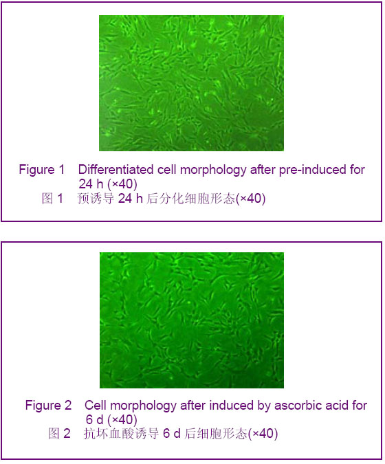
Figure 2 Cell morphology after induced by ascorbic acid for
6 d (×40)
图2 抗坏血酸诱导6 d后细胞形态(×40) Figure 2 Cell morphology after induced by ascorbic acid for 6 d (×40) 图2 抗坏血酸诱导6 d后细胞形态(×40)
全反式维甲酸+胶质细胞源性神经营养因子组:加入全反式维甲酸和胶质细胞源性神经营养因子1 h后可观察到扁平细胞的胞体向内收缩,呈圆形或椭圆形,并向周围长出较长的突起,5 h后分化为简单双极或大的多极细胞,胞体能伸展很长的突触,表现出典型的神经元样细胞形态,有立体感,折光性增强,见图3。
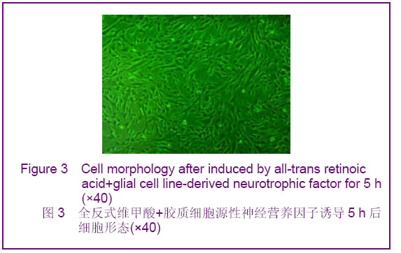
Figure 3 Cell morphology after induced by all-trans retinoic acid+glial cell line-derived neurotrophic factor for 5 h (×40) 图3 全反式维甲酸+胶质细胞源性神经营养因子诱导5 h后细胞形态(×40)
全反式维甲酸+胶质细胞源性神经营养因子+抗坏血酸组:0.5 h后即可观察到细胞胞质收缩,变小,呈不规则形、圆形及椭圆形,并逐渐向周围长出较长的突起,有立体感,折光性强,5 h后分化为简单单极、双极或多极细胞,极少数表现为锥形,胞体能伸展较长的突触,表现出典型的神经元形态,少数细胞周围有光晕,部分细胞交织,见图4。
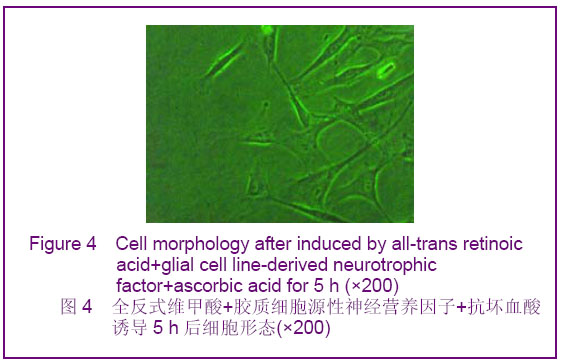
Figure 4 Cell morphology after induced by all-trans retinoic acid+glial cell line-derived neurotrophic factor+ascorbic acid for 5 h (×200) 图4 全反式维甲酸+胶质细胞源性神经营养因子+抗坏血酸诱导5 h后细胞形态(×200)
2.3 免疫细胞化学染色结果
神经细胞的鉴定:各诱导组均可见Nestin、神经元特异性烯醇化酶、胶质纤维酸性蛋白阳性细胞,表明人脐血间充质干细胞有多向分化潜能,具有神经细胞的基本特征,见图5-7。
Figure 5 Nestin positive cells (×200)图5 Nestin阳性细胞(×200) Figure 6 Neuron-specific enolase positive cells (×200) 图6 神经元特异性烯醇化酶阳性细胞(×200) Figure 7 Glial fibrillary acidic protein positive cells (×200) 图7 胶质纤维酸性蛋白阳性细胞(×200)
人脐血间充质干细胞向多巴胺能神经元的分化:各诱导组均可见酪氨酸羟化酶、多巴胺转运体、多巴胺受体D2阳性细胞,阳性细胞的胞浆呈棕黄色,胞核无表达,见图8-10。
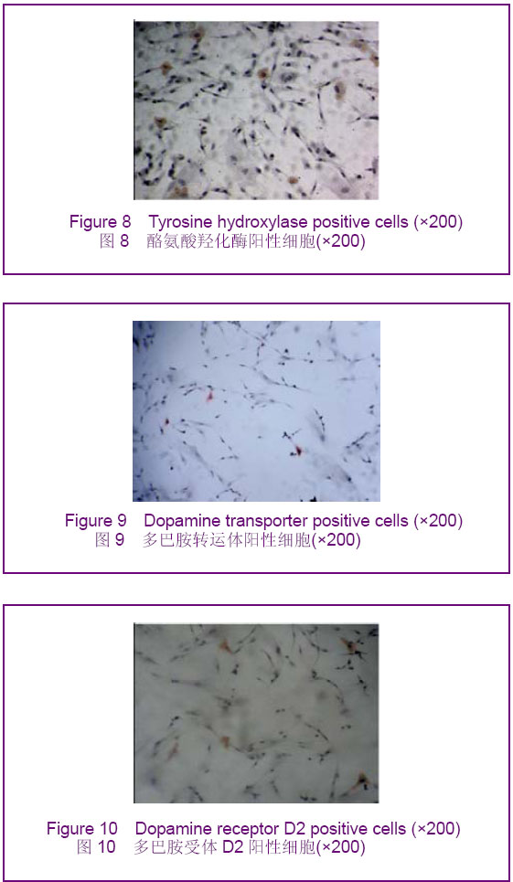
Figure 8 Tyrosine hydroxylase positive cells (×200)图8 酪氨酸羟化酶阳性细胞(×200)
Figure 9 Dopamine transporter positive cells (×200)图9 多巴胺转运体阳性细胞(×200)
Figure 10 Dopamine receptor D2 positive cells (×200)图10 多巴胺受体D2阳性细胞(×200)
抗坏血酸,全反式维甲酸 +胶质细胞源性神经营养因子,抗坏血酸+全反式维甲酸+胶质细胞源性神经营养因子各诱导组酪氨酸羟化酶阳性细胞率分别为(7.98±1.59)%,(13.50±1.45)%,(19.17±2.27)%。
多巴胺转运体阳性细胞率:抗坏血酸,全反式维甲酸 +胶质细胞源性神经营养因子,抗坏血酸+全反式维甲酸+胶质细胞源性神经营养因子各诱导组多巴胺转运体阳性细胞率分别为(6.22±1.62)%,(12.07±1.42)%,(17.61±1.45)%。
多巴胺受体D2阳性细胞率:抗坏血酸,全反式维甲酸+胶质细胞源性神经营养因子,抗坏血酸+全反式维甲酸+胶质细胞源性神经营养因子各诱导组多巴胺受体D2阳性细胞率分别为(0.22±0.08)%,(0.39±0.15)%,(0.48± 0.13)%。与对照组比较,各诱导组Nestin、神经元特异性烯醇化酶、胶质纤维酸性蛋白、酪氨酸羟化酶、多巴胺转运体、多巴胺受体D2阳性细胞率均明显升高(
P < 0.05)。与抗坏血酸组比较,全反式维甲酸+胶质细胞源性神经营养因子组和抗坏血酸+全反式维甲酸+胶质细胞源性神经营养因子组均明显升高(
P < 0.05),且抗坏血酸+全反式维甲酸+胶质细胞源性神经营养因子组升高幅度高于全反式维甲酸+胶质细胞源性神经营养因子组(
P < 0.05)。
2.4 Real Time RT-PCR检测结果 与对照组相比,各诱导组中酪氨酸羟化酶mRNA、多巴胺转运体 mRNA、多巴胺受体D2 mRNA的含量均有所增加,其中以抗坏血酸+全反式维甲酸+胶质细胞源性神经营养因子组酪氨酸羟化酶mRNA、多巴胺转运体mRNA、多巴胺受体D2mRNA的含量增加最为明显,全反式维甲酸+胶质细胞源性神经营养因子组次之,抗坏血酸组单独诱导较弱,差异有显著性意义(
P < 0.05),见表1。
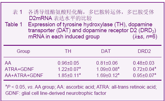
表1 各诱导组酪氨酸羟化酶,多巴胺转运体,多巴胺受体D2mRNA表达水平的比较
Table 1 Expression of tyrosine hydroxylase (TH), dopamine transporter (DAT) and dopamine receptor D2 (DRD2) mRNA in each induced group (x±s, n=6)


