| [1]Birbrair A, Zhang T, Wang Z M, et al. Type-2 pericytes participate in normal and tumoral angiogenesis. Am J Physiol Cell Physiol.2014;307(1):C25-C38.[2]Kim B, Lee Y, Yoo H, et al. Anti-angiogenic activity of thienopyridine derivative LCB03-0110 by targeting VEGFR-2 and JAK/STAT3 Signalling. Exp Dermatol. 2015;24(7):503-509.[3]王丽,李录英,陆齐,等. CD73在乳腺癌血管新生中的作用[J].中国病理生理杂志,2015,12(10):1843.[4]Li S,Haigh K,Haigh JJ,et al.Endothelial VEGF sculpts cortical cytoarchitecture.J Neurosci.2013;33(37):14809-14815.[5]江克华,董自强,宋兴福,等.PI3K/PTEN信号通路与肿瘤血管形成关系的研究进展[J].实用医学杂志,2011,27(22):4169-4170.[6]李景,付焱,肖光雄,等.源于乳腺癌干细胞的内皮细胞参与肿瘤血管形成的实验研究[J].医学研究生学报, 2016,29(11): 1191-1196.[7]陈银涛,于秉治,武迪迪. PI3K/Akt/mTOR信号通路及临床相关肿瘤抑制剂[J].中国生物化学与分子生物学报, 2014,30(10):949-956.[8]廖明娟,陈红风.PI3K/Akt/mTOR信号通路抑制剂在乳腺癌中的研究进展[J]. 中华肿瘤防治杂志,2012,19(3):230-234.[9]王法斌,李京波. 他汀类药物对血管形成的作用及分子机制[J]. 实用医学杂志,2012,28(23):4018-4020.[10]许小兵,郭美霞,金鑫鑫,等. 塞来昔布抑制血管内皮细胞迁移的实验研究[J]. 医学研究生学报,2017,30(6):601-605.[11]王宇霆,周海燕,吕小翠.白藜芦醇对过氧化氢氧化应激损伤人脐静脉血管内皮细胞的保护作用[J].中华医学杂志, 2013,93(15): 1174-1177.[12]马丽娜,陈北冬,赵艳阳,等.银杏内酯B对内皮细胞的保护作用及分子机制研究[J].中国药理学通报,2013,29(2):189-193.[13]Ghosh CC, Mukherjee A, David S, et al. Angiopoietin-1 Requires Oxidant Signaling through p47phox to Promote Endothelial Barrier Defense.PLoS One. 2015;10(3): e119577.[14]周政荣,丁丽娜.冠心病合并糖尿病应用奥美沙坦酯对患者血管内皮细胞功能的影响作用[J].北方药学,2018,15(11):29-30.[15]王志钢,吴应积,旭日干. mTOR信号通路与细胞生长调控[J]. 生物物理学报,2007,23(5):333-342.[16]高阳,田树旭,常小丽,等.雷帕霉素靶蛋白的蛋白信号通路及其与代谢性疾病相关性[J].医学研究生学报,2010,23(2):196-201.[17]Chung AS,Lee J, Ferrara N. Targeting the tumour vasculature: insights from physiological angiogenesis. Nat Rev Cancer. 2010;10(7):505-514.[18]Zeng Q,Wu Z,Duan H,et al.Impaired tumor angiogenesis and VEGF-induced pathway in endothelial CD146 knockout mice. Protein Cell.2014;5(6):445-456.[19]Huang B,Wang Y,Wang W,et al. mTORC1 Prevents Preosteoblast Differentiation through the Notch Signaling Pathway. PLOS Genetics.2015;11(8):400-426.[20]Ma A, Wang L, Gao Y, et al. Tsc1 deficiency-mediated mTOR hyperactivation in vascular endothelial cells causes angiogenesis defects and embryonic lethality. Human Molecular Genetics.2014,23(3):693-705.[21]Duan S, Shafi JRS, Kuchay S, et al. mTOR Generates an Auto-Amplification Loop by Triggering the bTrCP- and CK1a-Dependent Degradation of DEPTOR. 2011:44: 317-324.[22]Zhan JK, Wang YJ, Wang Y, et al. Adiponectin attenuates the osteoblastic differentiation of vascular smooth muscle cells through the AMPK/mTOR pathway. Exp Cell Res.2014; 323(2):352-358.[23]Laplante M, Horvat S, Festuccia WT, et al. DEPTOR Cell-Autonomously Promotes Adipogenesis, and Its Expression Is Associated with Obesity. Cell Metab. 2012; 16(2):202-212.[24]Kazi AA,Hong-Brown L,Lang SM, et al. Deptor knockdown enhances mTOR Activity and protein synthesis in myocytes and ameliorates disuse muscle atrophy. Mol Med.2011; 17(9-10):925-936.[25]Bruneau S,Nakayama H,Woda CB,et al.DEPTOR regulates vascular endothelial cell activation and proinflammatory and angiogenic responses. Blood. 2013;122(10):1833-1842.[26]Liu M, Wilk SA, Wang A, et al. Resveratrol inhibits mTOR signaling by promoting the interaction between mTOR and DEPTOR.J Biol Chem.2010;285(47):36387-36394.[27]赖舒畅,邱鸿,王肖,等.条件性胰岛β细胞DEPTOR基因敲除小鼠构建及鉴定[J].实用医学杂志,2018,34(4):552-555.[28]王志蕊,刘西梅,周荆荣,等. Cre-LoxP重组系统删除内源性选择标记基因的效能评价[J]. 中国生物化学与分子生物学报, 2014, 30(2):194-201.[29]吴壮,徐军.Cre/Loxp位点重组酶系统在疾病动物模型建立中的应用[J].国外医学(呼吸系统分册),2004,24(4):254-256.[30]任荣荣,王英伟. Cre/Loxp系统的应用及进展[J].广州医学院学报,2008,36(2):78-80.[31]Hong J,Liu R,Chen L,et al. Conditional knockout of tissue factor pathway inhibitor 2 in vascular endothelial cells accelerates atherosclerotic plaque development in mice. Thromb Res.2016;137:148-156.[32]桑璐,郭英,任立明,等.利用Cre/lox重组系统在转基因动物中筛选高效表达外源基因的"友好"基因座[J]. 高技术通讯,2004, 14(12):43-49.[33]Campochiaro PA, Sophie R, Tolentino M, et al. Treatment of diabetic macular edema with an inhibitor of vascular endothelial-protein tyrosine phosphatase that activates tie2.Ophthalmology.2015;122(3):545-554.[34]Sarkar K, Rey S, Zhang X, et al. Tie2-dependent knockout of HIF-1 impairs burn wound vascularization and homing of bone marrow-derived angiogenic cells. Cardiovascular Res. 2011;93(1):162-169.[35]Kawakami Y, Ii M, Matsumoto T, et al. SDF-1/CXCR4 Axis in Tie2-Lineage Cells Including Endothelial Progenitor Cells Contributes to Bone Fracture Healing. J Bone Miner Res. 2015;30(1):95-105.[36]Catena V,Fanciulli M.Deptor: not only a mTOR inhibitor. J Exp Clin Cancer Res. 2017;36(1):12.[37]Ji YM, Zhou XF, Zhang J, et al. DEPTOR suppresses the progression of esophageal squamous cell carcinoma and predicts poor prognosis.Oncotarget.2016;7(12):14188-14198.[38]Chen L, Liu T, Tu Y, et al. Cul1 promotes melanoma cell proliferation by promoting DEPTOR degradation and enhancing cap-dependent translation. Oncol Rep.2016; 35(2):1049-1056.[39]Caron A,Mouchiroud M,Gautier N,et al.Loss of hepatic DEPTOR alters the metabolic transition to fasting. Molecular Metabolism.2017;6(5):447-458.[40]Dong X,Wang L, Han Z, et al. Different functions of DEPTOR in modulating sensitivity to chemotherapy for esophageal squamous cell carcinoma. Exp Cell Res. 2017;353(1):35-45.[41]Zhou X,Guo J,Ji Y,et al. Reciprocal Negative Regulation between EGFR and DEPTOR Plays an Important Role in the Progression of Lung Adenocarcinoma. Mol Cancer Res. 2016; 14(5):448-457.[42]Srinivas KP, Viji R, Dan VM, et al. DEPTOR promotes survival of cervical squamous cell carcinoma cells and its silencing induces apoptosis through downregulating PI3K/AKT and by up-regulating p38 MAP kinase. Oncotarget. 2016;7(17): 24154-24171.[43]Agrawal P,Reynolds J,Chew S,et al.DEPTOR is a stemness factor that regulates pluripotency of embryonic stem cells. J Biol Chem.2014;289(46):31818-31826.[44]González-Terán B, López JA, Rodríguez E, et al. p38γ and δ promote heart hypertrophy by targeting the mTOR-inhibitory protein DEPTOR for degradation. Nat Commun. 2016;7: 10477.[45]Ding Y,Shan L,Nai W,et al.DEPTOR Deficiency-Mediated mTORc1 Hyperactivation in Vascular Endothelial Cells Promotes Angiogenesis. Cell Physiol Biochem. 2018;46(2): 520-531 [46]Xu N,Guan S,Chen Z,et al.The alteration of protein prenylation induces cardiomyocyte hypertrophy through Rheb-mTORC1 signalling and leads to chronic heart failure. J Pathol.2015;235(5):672-685.[47]Nitta N, Nakasu S, Shima A, et al. mTORC1 signaling in primary central nervous system lymphoma. Surg Neurol Int.2016;7(Suppl 17):S475-S480.[48]Lorbeer R,Baumeister SE,Dorr M,et al.Angiopoietin-2, its soluble receptor Tie-2 and subclinical cardiovascular disease in a population-based sample. Heart.2015;101(3):178-184.[49]Kisanuki YY, Hammer RE, Miyazaki J,et al.Tie2-Cre transgenic mice: a new model for endothelial cell-lineage analysis in vivo. Dev Biol.2001;230(2):230-242.[50]Barry DM, Xu K,Meadows SM, et al. Cdc42 is required for cytoskeletal support of endothelial cell adhesion during blood vessel formation in mice. Development.2015;142(17): 3058-3070.[51]Ohashi K,Ouchi N,Higuchi A, et al. LKB1 deficiency in Tie2-Cre-expressing cells impairs ischemia-induced angiogenesis.J Biol Chem.2010;285(29):22291-22298.[52]丁燕,赖丽芬,奈文青,等.血管内皮细胞敲除Rheb基因杂合子小鼠模型建立及意义[J].山东医药,2017,57(36):34-36.[53]李娇,陈永文,张庆镐. CD1d基因敲除小鼠拮抗DSS诱导结肠炎[J].免疫学杂志,2018,34(9):759-764. |
.jpg)
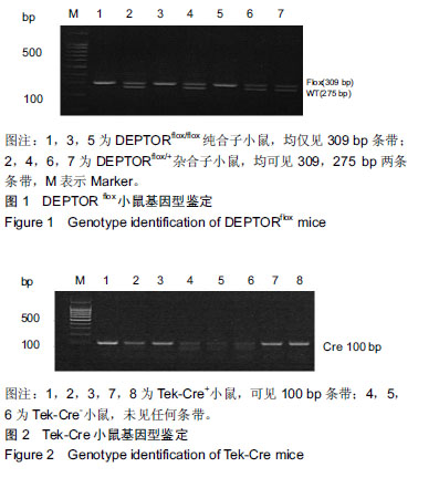
.jpg)
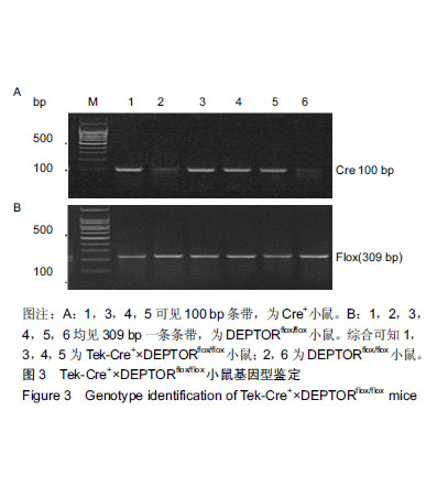
.jpg) #br#
文题释义:#br#
DEPTOR:含有DEP结构域的mTOR相互作用蛋白(DEPTOR),也称为含有DEP结构域的蛋白6(DEPDC6),是人类中由DEPTOR基因编码的蛋白质。DEPTOR在调节血管内皮细胞活化,并且在体外促炎和血管生成反应中发挥重要作用。#br#
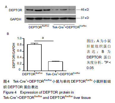
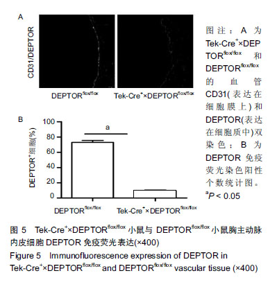
血管内皮细胞DEPTOR基因敲除模型建立:利用Cre/loxp技术建立小鼠血管内皮细胞DEPTOR基因敲除模型,利用基因鉴定和蛋白水平验证敲除效果,初步观察敲除小鼠表型。
#br#
文题释义:#br#
DEPTOR:含有DEP结构域的mTOR相互作用蛋白(DEPTOR),也称为含有DEP结构域的蛋白6(DEPDC6),是人类中由DEPTOR基因编码的蛋白质。DEPTOR在调节血管内皮细胞活化,并且在体外促炎和血管生成反应中发挥重要作用。#br#
血管内皮细胞DEPTOR基因敲除模型建立:利用Cre/loxp技术建立小鼠血管内皮细胞DEPTOR基因敲除模型,利用基因鉴定和蛋白水平验证敲除效果,初步观察敲除小鼠表型。