中国组织工程研究 ›› 2018, Vol. 22 ›› Issue (25): 3969-3974.doi: 10.3969/j.issn.2095-4344.0955

• 脂肪干细胞 adipose-derived stem cells • 上一篇    下一篇

自体脂肪干细胞、壳聚糖/胶原蛋白支架材料及GDF-5复合物移植修复兔软骨缺损

王 征1,于 会1,于 尧1,吴柏霖2   

  1. 1沈阳医学院附属中心医院手外科,辽宁省沈阳市 110024;2山东大学,山东省济南市 250100
  • 修回日期:2018-05-19 出版日期:2018-09-08 发布日期:2018-09-08
  • 通讯作者: 王征,沈阳医学院附属中心医院手外科,辽宁省沈阳市 110024
  • 作者简介:王征,硕士,主治医师。
  • 基金资助:

    沈阳医学院青年基金(20142050);沈阳市卫生和计划生育委员会科研基金

Repairing articular cartilage defects in rabbits by adipose-derived stem cells combined with chitosan/collagen scaffold and GDF-5

Wang Zheng1, Yu Hui1, Yu Yao1, Wu Bo-lin2   

  1. 1Department of Hand Surgery, Central Hospital Affiliated to Shenyang Medical College, Shenyang 110024, Liaoning Province, China; 2Shandong University, Jinan 250100, Shandong Province, China
  • Revised:2018-05-19 Online:2018-09-08 Published:2018-09-08
  • Contact: Wang Zheng, Department of Hand Surgery, Central Hospital Affiliated to Shenyang Medical College, Shenyang 110024, Liaoning Province, China
  • About author:Wang Zheng, Master, Attending physician, Department of Hand Surgery, Central Hospital Affiliated to Shenyang Medical College, Shenyang 110024, Liaoning Province, China
  • Supported by:

    the Youth Foundation of Shenyang Medical College, No. 20142050; the Fund of Shenyang Health and Family Planning Commission

摘要:

文章快速阅读:

文题释义:
GDF-5因子:
又称为软骨形态发生蛋白1,可促进早期软骨和关节形成。
GDF-5复合物:应用紫外线交联法以1∶3比例制备构建壳聚糖/胶原蛋白复合支架载体。将标记后的兔脂肪干细胞种植到支架载体上共同培养形成复合物,再加入GDF-5滴注浓度为100 μg/L即构建脂肪干细胞+壳聚糖/胶原蛋白支架+GDF-5复合物。


摘要
背景:
GDF-5可诱导人脂肪干细胞分化为软骨样细胞,壳聚糖/胶原蛋白支架在软骨组织损伤修复中应用广泛。
目的:利用兔的自体脂肪干细胞复合壳聚糖/胶原蛋白支架材料加入GDF-5修复兔关节软骨缺损,观察其修复效果。
方法:取日本大耳兔皮下脂肪组织提取脂肪干细胞体外培养、纯化、扩增,并BrdU体外标记,应用紫外线交联法以1∶3比例构建壳聚糖/胶原蛋白复合支架载体,将标记后的兔脂肪干细胞种植到支架载体上共同培养形成复合物,以手术方式造成兔膝关节软骨直径为5 mm,深3 mm的圆柱形缺损,建立膝关节软骨缺损模型兔,随机分为4组,复合物组:脂肪干细胞+壳聚糖/胶原蛋白支架+GDF-5复合物植入干预;支架组:单纯植入壳聚糖/胶原蛋白支架;脂肪干细胞组:单纯植入脂肪干细胞;假手术组:单纯钻孔不作处理。各组干预4,8,12周时取材,行组织学检测、软骨Ⅱ型胶原免疫组织化学染色及BrdU免疫荧光染色。
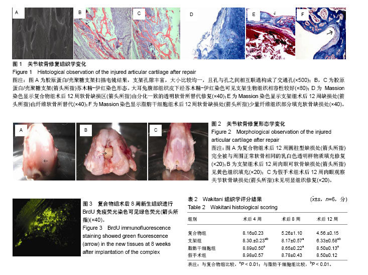
结果与结论:①术后12周,与其他3组相比,复合物组圆柱型缺损处完全被与周围正常软骨相同的乳白色透明样物质填充修复,软骨缺损区域由分化一致的透明软骨所替代修复,Wakitani评分最低(P < 0.01);②Ⅱ型胶原免疫组化染色及BrdU免疫荧光染色:复合物组与术后4,8,12周免疫组化染色可见修复区组织Ⅱ型胶原染色胞浆内呈淡黄色阳性表达,修复区新生组织可见BrdU绿色荧光表达;③结果证实,自体脂肪干细胞+壳聚糖/胶原蛋白支架+GDF-5复合物移植修复兔膝关节软骨缺损效果较好。

中国组织工程研究杂志出版内容重点:干细胞;骨髓干细胞;造血干细胞;脂肪干细胞;肿瘤干细胞;胚胎干细胞;脐带脐血干细胞;干细胞诱导;干细胞分化;组织工程
ORCID:
0000-0003-2061-3210(王征)

关键词: 壳聚糖, 胶原蛋白, 支架;组织工程, 脂肪干细胞, GDF-5, 兔, 软骨缺损, 损伤修复, 自体

Abstract:

BACKGROUND: GDF-5 can induce the differentiation of human adipose-derived stem cells into chondrocyte-like cells, and chitosan/collagen scaffolds are widely used in cartilage tissue repair.
OBJECTIVE: To repair rabbit articular cartilage defects by using rabbit autologous adipose-derived stem cells combined with chitosan/collagen scaffold and GDF-5 and to observe the repair effect.
METHODS: Adipose-derived stem cells isolated from subcutaneous adipose tissues of Japanese rabbits were cultured, purified, amplified in vitro, and labeled with BrdU in vitro. The chitosan/collagen composite scaffold was prepared in a ratio of 1:3 using ultraviolet cross-linking method. Then labeled adipose-derived stem cells were seeded onto the scaffold and cultured to form complexes. A cylindrical defect (5 mm in diameter, 3 mm in depth) in the knee articular cartilage was surgically made in each rabbit, and these rabbit models were thereafter randomly divided into four groups. Composite group: adipose-derived stem cells+chitosan/collagen scaffold+GDF-5 complex implantation; stent group: simple implantation of chitosan/collagen scaffold; cell group: simple implantation of adipose-derived stem cells; and sham operation group: simple drilling without treatment. All the rabbits were intervened for 4, 8, and 12 weeks. Meanwhile, histological detection, type II collagen immunohistochemical staining and BrdU mmunofluorescence staining were performed.
RESULTS AND CONCLUSION: (1) At 12 weeks after surgery, the cylindrical defect of the composite group was completely filled with the same milky white transparent substance as the surrounding normal cartilage, and the cartilage defect area was replaced by differentiated hyaline cartilage. Of all the groups, Wakitani score was the lowest in the composite group (P < 0.01). (2) Type II collagen immunohistochemical staining and BrdU immunofluorescence staining: in the composite group, the type II collagen stained cytoplasm in the repaired area was detected by immunohistochemical staining at 4, 8, and 12 weeks after surgery. The expression of BrdU in the new tissues of the repaired area was positive. These results confirm that autologous adipose-derived stem cells+chitosan/collagen scaffold+GDF-5 complex grafting has a good effect on the repair of rabbit knee cartilage defects.

中国组织工程研究杂志出版内容重点:干细胞;骨髓干细胞;造血干细胞;脂肪干细胞;肿瘤干细胞;胚胎干细胞;脐带脐血干细胞;干细胞诱导;干细胞分化;组织工程

Key words: Stem Cells, Tissue Engineering, Chitosan, Stents

中图分类号: