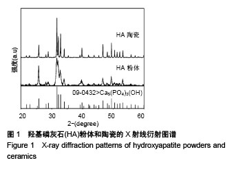
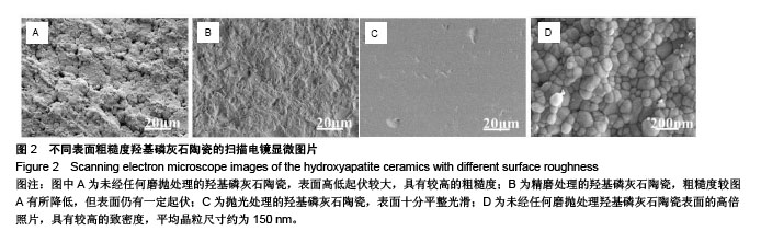
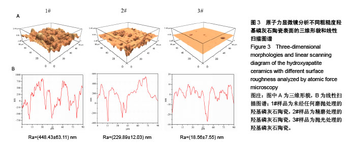
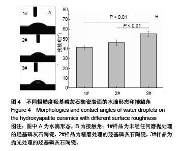
| [1] Yang XY,Chen LH,Li Y,et al.Hierarchically porous materials: synthesis strategies and structure design.Chem Soc Rev. 2017;46:481-558.[2] Zadpoor AA.Bone tissue regeneration: the role of scaffold geometry. Biomater Sci-Uk. 2015;3:231-245.[3] Tang Z,Li X,Tan Y,et al.The material and biological characteristics of osteoinductive calcium phosphate ceramics.Regen Biomater.2018; 5:43-59.[4] Zhang K,Fan Y,Dunne N,et al.Effect of microporosity on scaffolds for bone tissue engineering. Regenerative Biomaterials.2018;5:115-124.[5] Hong YL,Fan HS,Li B,et al.Fabrication, biological effects, and medical applications of calcium phosphate nanoceramics.Mat Sci Eng R. 2010; 70:225-242.[6] Ranella A,Barberoglou M,Bakogianni S,et al.Tuning cell adhesion by controlling the roughness and wettability of 3D micro/nano silicon structures.Acta Biomater. 2010;6:2711-2720.[7] Rosales-Leal JI,Rodriguez-Valverde MA,Mazzaglia G,et al.Effect of roughness, wettability and morphology of engineered titanium surfaces on osteoblast-like cell adhesion.Colloid Surface A.2010;365:222-229.[8] Dee KC,Puleo DA,Bizios R.Protein-surface interactions. In: Dee KC, Puleo DA, Bizios R, editors. An introduction to tissue-biomaterial interactions.New York:John Wiley & Sons, 2002:37-52.[9] Wang J,Zhu Y,Wang M,et al.Fabrication and preliminary biological evaluation of a highly porous biphasic calcium phosphate scaffold with nano-hydroxyapatite surface coating.Ceram Int. 2018;44:1304-1311.[10] Bohner M,Loosli Y,Baroud G,et al.Commentary: Deciphering the link between architecture and biological response of a bone graft substitute. Acta Biomater.2011;7:478-484.[11] Best SM,Porter AE,Thian ES,et al.Bioceramics: Past, present and for the future.J Eur Ceram Soc.2008;28:1319-1327.[12] Dorozhkin SV.Nanodimensional and Nanocrystalline Apatites and Other Calcium Orthophosphates in Biomedical Engineering, Biology and Medicine.Materials. 2009;2:1975-2045.[13] Tripathi G,Basu B.A porous hydroxyapatite scaffold for bone tissue engineering: Physico-mechanical and biological evaluations.Ceram Int.2012;38:341-349.[14] Yang PP,Quan ZW,Li CX,et al.Bioactive, luminescent and mesoporous europium-doped hydroxyapatite as a drug carrier.Biomaterials. 2008; 29:4341-4347.[15] Civelek AC,Pacheco EM,Natarajan TK,et al.Quantitative Measurement of Vascularization and Vascular Ingrowth Rate of Coralline Hydroxyapatite Ocular Implant by Tc-99m Mdp Bone Imaging. Clin Nucl Med.1995;20:779-787.[16] Cowper TR.Hydroxyapatite Motility Implants in Ocular Prosthetics.J Prosthet Dent. 1995;73:267-273.[17] Bose S,Dasgupta S,Tarafder S,et al.Microwave-processed nanocrystalline hydroxyapatite: Simultaneous enhancement of mechanical and biological properties.Acta Biomater. 2010;6:3782-3790.[18] Lin KL,Chen L,Chang J.Fabrication of Dense Hydroxyapatite Nanobioceramics with Enhanced Mechanical Properties via Two-Step Sintering Process.Int J Appl Ceram Tec.2012;9:479-485.[19] Adeleke SA,Ramesh S,Bushroa AR,et al.The properties of hydroxyapatite ceramic coatings produced by plasma electrolytic oxidation. Ceram Int.2018;44:1802-1811.[20] Machado GC,Garcia-Tunon E,Bell RV,et al.Calcium phosphate substrates with emulsion-derived roughness: Processing, characterisation and interaction with human mesenchymal stem cells.J Eur Ceram Soc.2018;38:949-961.[21] Roy M,Fielding GA,Beyenal H,et al.Mechanical, In vitro Antimicrobial, and Biological Properties of Plasma-Sprayed Silver-Doped Hydroxyapatite Coating.Acs Appl Mater Inter. 2012;4:1341-1349.[22] Heimann RB.Plasma-Sprayed Hydroxylapatite-Based Coatings: Chemical, Mechanical, Microstructural,and Biomedical Properties.J Therm Spray Techn.2016;25:827-850.[23] Zhang XD,Yuan HP,de Groot K.Calcium phosphate biomaterials with intrinsic osteoinductivity. The 6th world biomaterials congress.Hawaii, USA,2000:15-20.[24] Tang XM,Huang K,Dai J,et al.Influences of surface treatments with abrasive paper and sand-blasting on surface morphology, hydrophilicity, mineralization and osteoblasts behaviors of n-CS/PK composite.Sci Rep.2017;7(1):568. [25] Fujibayashi S,Neo M,Kim HM,et al.Osteoinduction of porous bioactive titanium metal.Biomaterials.2004;25:443-450.[26] Del Fabbro M,Taschieri S,Canciani E,et al.Osseointegration of Titanium Implants With Different Rough Surfaces: A Histologic and Histomorphometric Study in an Adult Minipig Model.Implant Dent. 2017;26:357-366.[27] Lin KL,Xia LG,Gan JB,et al.Tailoring the Nanostructured Surfaces of Hydroxyapatite Bioceramics to Promote Protein Adsorption, Osteoblast Growth, and Osteogenic Differentiation.Acs Appl Mater Inter.2013;5:8008-8017.[28] Hayashi K,Inadome T,Tsumura H,et al.Effect of surface roughness of hydroxyapatite-coated titanium on the bone-implant interface shear strength.Biomaterials. 1994;15:1187-1191.[29] Puleo DA,Nanci A.Understanding and controlling the bone-implant interface.Biomaterials. 1999;20:2311-2321.[30] Zhu XD,Zhang HJ,Fan HS,et al.Effect of phase composition and microstructure of calcium phosphate ceramic particles on protein adsorption.Acta Biomater.2010;6:1536-1541.[31] Zhu XD,Fan HS,Xiao YM,et al.Effect of surface structure on protein adsorption to biphasic calcium-phosphate ceramics in vitro and in vivo.Acta Biomater.2009;5:1311-1318.[32] Zhu XD,Zhang HJ,Li DX,et al.Study on the enhanced protein adsorption of microwave sintered hydroxyapatite nanoceramic particles: Role of microstructure.J Biomed Mater Res B Appl Biomater.2012;100(2): 516-523.[33] Hennessy KM,Clem WC,Phipps MC,et al.The effect of RGD peptides on osseointegration of hydroxyapatite biomaterials.Biomaterials. 2008; 29:3075-3083.[34] Deligianni DD,Katsala N,Ladas S,et al.Effect of surface roughness of the titanium alloy Ti-6Al-4V on human bone marrow cell response and on protein adsorption.Biomaterials. 2001;22:1241-1251.[35] Deligianni DD,Katsala ND,Koutsoukos PG,et al.Effect of surface roughness of hydroxyapatite on human bone marrow cell adhesion, proliferation, differentiation and detachment strength. Biomaterials. 2001;22:87-96.[36] dos Santos EA,Farina M,Soares GA,et al.Chemical and topographical influence of hydroxyapatite and beta-tricalcium phosphate surfaces on human osteoblastic cell behavior.J Biomed Mater Res A.2009;89A:510-520. |
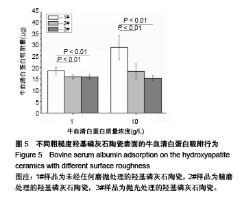
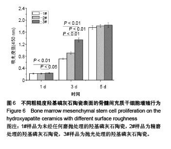
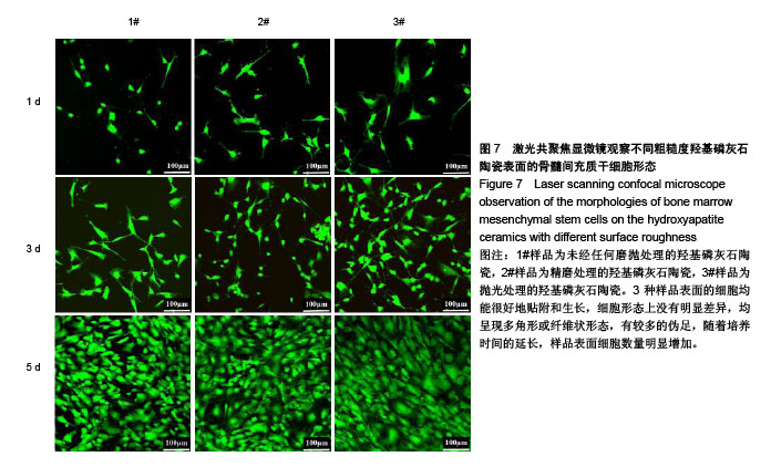
.jpg)
.jpg)