中国组织工程研究 ›› 2018, Vol. 22 ›› Issue (28): 4457-4462.doi: 10.3969/j.issn.2095-4344.0837

• 软骨组织构建 cartilage tissue construction • 上一篇    下一篇

成年关节软骨碎块化后MT1-MMP表达与软骨细胞迁移的关系

戴  祝1,彭嘉斌1,2,廖  瑛1,符得红1,雷运亮1,黎  洲1   

  1. 1南华大学附属第一医院骨科一病区,湖南省衡阳市  421000;2广东省中医院珠海医院骨五科,广东省珠海市  519000
  • 收稿日期:2018-01-26 出版日期:2018-10-08 发布日期:2018-10-08
  • 通讯作者: 彭嘉斌,硕士,医师,南华大学附属第一医院骨科一病区,湖南省衡阳市 421000;广东省中医院珠海医院骨五科,广东省珠海市 519000
  • 作者简介:戴祝,男,1980年生,湖南省长沙市人,汉族,2013年中南大学毕业,博士,副主任医师,主要从事关节与运动医学研究。
  • 基金资助:

    国家自然科学基金资助项目(81501886);湖南省自然科学基金资助项目(2015JJ3102)

Relationship of membrane type-1 matrix metalloproteinase expression and chondrocyte migration in adult minced articular cartilage

Dai Zhu1, Peng Jia-bin1, 2, Liao Ying1, Fu De-hong1, Lei Yun-liang1, Li Zhou1   

  1. 1First Ward of Orthopedics, the First Affiliated Hospital of University of South China, Hengyang 421000, Hunan Province, China; 2Fifth Department of Orthopedics, Guangdong Hospital of Traditional Chinese Medicine Zhuhai Branch, Zhuhai 519000, Guangdong Province, China
  • Received:2018-01-26 Online:2018-10-08 Published:2018-10-08
  • Contact: Peng Jia-bin, Master, Physician, First Ward of Orthopedics, the First Affiliated Hospital of University of South China, Hengyang 421000, Hunan Province, China; Fifth Department of Orthopedics, Guangdong Hospital of Traditional Chinese Medicine Zhuhai Branch, Zhuhai 519000, Guangdong Province, China
  • About author:Dai Zhu, M.D., Associate chief physician, First Ward of Orthopedics, the First Affiliated Hospital of University of South China, Hengyang 421000, Hunan Province, China
  • Supported by:

    the National Natural Science Foundation of China, No. 81501886; the Natural Science Foundation of Hunan Province, No. 2015JJ3102

摘要:

文章快速阅读:
文题释义:
细胞迁移:也称为细胞爬行、细胞移动或细胞运动,是指细胞在接收到迁移信号或感受到某些物质的梯度后而产生的移动。细胞迁移为细胞头部伪足的延伸、新的黏附建立、细胞体尾部收缩在时空上的交替过程。细胞迁移是正常细胞的基本功能之一,是机体正常生长发育的生理过程,也是活细胞普遍存在的一种运动形式。胚胎发育、血管生成、伤口愈合、免疫反应、炎症反应、动脉粥样硬化、癌症转移等过程中都涉及细胞迁移。
MT1-MMP:是基质金属蛋白酶(matrix metalloproteinase,MMP)家族的新成员,基因定位于14q11,长度超过10 kb,包括10个外显子和9个内含子,编码582个氨基酸。MT1-MMP主要作用是介导细胞迁移。
摘要
背景:
许多课题研究已基本证实,关节软骨碎块化处理后,软骨损伤区域的修复效应增强,但对于其具体机制仍不清楚。
目的:研究成年关节软骨经碎块化处理后,MT1-MMP表达与软骨细胞迁移的关系,进一步探讨软骨经碎块化后,修复效应增强的机制。
方法:通过无菌操作从78只成年家兔双后腿膝关节(156膝)获取碎块关节软骨颗粒(约1 mm×1 mm×1 mm)和块状软骨(直径约5 mm,厚度约2 mm),复合明胶海绵碎屑及纤维蛋白胶制作体外培养模型。分组:碎块组和整块组均采用普通细胞培养液进行培养;MT1-MMP抑制剂组:采用添加外源性MT1-MMP抑制剂进行干预培养;各组经体外培养2,4,6周进行激光共聚焦显微镜观察细胞迁移,苏木精-伊红染色切片并进行组织半定量评分、碎块组与整块组标本进行MT1-MMP免疫组织化学检测并计算MT1-MMP阳性细胞百分率。
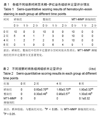
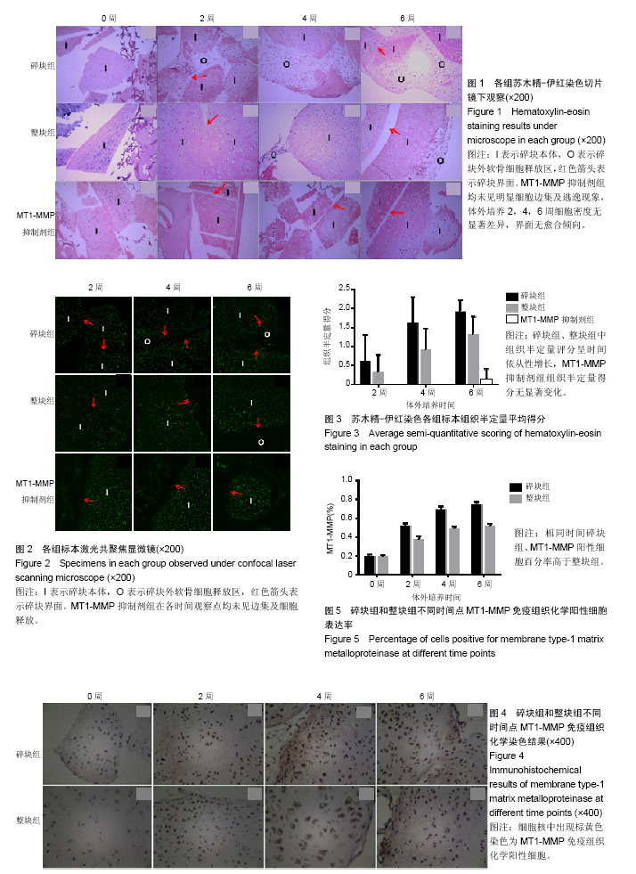
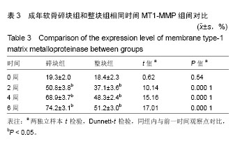
结果与结论:①激光共聚焦显微镜观察见碎块组、整块组经体外培养2,4周均可见软骨细胞边集现象,碎块组显著,且4,6周时可见大量软骨细胞释放;MT1-MMP抑制剂组在各时间观察点均未见边集及细胞释放;②组织半定量评分与MT1-MMP阳性细胞表达率在碎块组、整块组中随体外培养时间延长而增长,相同培养时间,碎块组得分及MT1-MMP阳性细胞百分率高于整块组;MT1-MMP抑制剂组均未见细胞边缘聚集及碎块愈合现象;③结果表明,成年家兔关节软骨经碎块化处理后其修复效应增强;同时成年家兔关节软骨经碎块处理后,MT1-MMP表达增高,软骨细胞迁移能力增强;抑制MT1-MMP功能,能有效抑制软骨细胞迁移及阻碍碎块愈合,在一定程度上可推测,MT1-MMP的表达增强是碎块化软骨损伤修复效应增强的可能机制。

中国组织工程研究杂志出版内容重点:组织构建;骨细胞;软骨细胞;细胞培养;成纤维细胞;血管内皮细胞;骨质疏松组织工程
ORCID: 0000-0002-7505-3858(戴祝)

关键词: 关节软骨, 软骨碎块, 细胞迁移, MT1-MMP, 组织构建

Abstract:

BACKGROUND: Minced articular cartilage has been shown to be helpful for cartilage repair, but the underlying mechanism remains unclear.
OBJECTIVE: To observe the association of expression level of membrane type-1 matrix metalloproteinase (MT1-MMP) with chondrocyte migration, and to further research the underlying mechanism.
METHODS: Massive cartilage (5 mm of diameter, and 2 mm of thickness) and articular cartilage fragments (about 1 mm3) were obtained from 78 adult rabbit keen joints under sterile condition, and then planted into fibrin glue-gelatin sponge composite scaffolds. Group A: cartilage fragments cultured in vitro with normal culture medium; group B: monoblock cartilage cultured in vitro with normal culture medium; group C: cartilage fragments cultured in vitro with culture medium containing inhibitor of MT1-MMP (NSC402050). The specimens were taken out respectively from each group after culture for 2, 4 and 6 weeks, for observing the chondrocyte migration under confocal laser scanning microscope, hematoxylin-eosin staining and histological semi-quantitative scoring. The expression level of MT1-MMP was detected by immunohistochemistry in the groups A and B, and the percentage of cells positive for MT1-MMP was calculated.
RESULTS AND CONCLUSION: Laser confocal microscope results showed that chondrocytes gathered at the margin of tissue after 2 and 4 weeks of in vitro culture in the groups A and B, especially the group A, and at the same time, there were numerous chondrocytes. Group C showed no chondrocyte gathered at the margin of tissue and there was no cell release. Semi-quantitative score and the percentage of cells positive for MT1-MMP in the groups A and B were increased with time in vitro, and the scores and MT1-MMP positive cells in the group A were higher than those in the group B. The semi-quantitative score in the group C was closed to 0. These results suggest that the minced articular cartilage can promote cartilage repair via up-regulating the expression level of MT1-MMP and increasing the migration of chondrocytes.

中国组织工程研究杂志出版内容重点:组织构建;骨细胞;软骨细胞;细胞培养;成纤维细胞;血管内皮细胞;骨质疏松组织工程

Key words: Joints, Chondrocytes, Cell Movement, Matrix Metalloproteinases, Tissue Engineering

中图分类号: