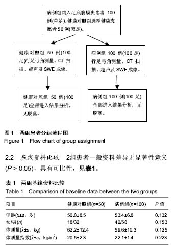
| [1] Oktem H, Calgüner E, Erdo?an D, et al. Age-related changes in light microscopy with Sirius red technique in rat achilles tendon J Eklem Hastalik Cerrahisi.2010;21(1): 50-55.[2] 毛宾尧,俞光荣. 踝足外科学[M].2版.北京:科学出版社, 2007.[3] Rathleff MS, Moelgaard C, Lykkegaard Oleasen J. Intra-and interobserver reliability of quantitative ultrasound measurement of the plantar fascia. J Clin Ultrasound. 2011;39(16): 128-134.[4] Kane D, Balint PV, Sturrock R, et al. Musculoskeletal ultrasound a state of the art review in rheumatology. Part 1: Current controversies and issues in the development of musculoskeletal ultrasound in rheumatology. J Rheumatology (Oxford).2004;43(4): 823-828.[5] Ozdemir H,Yilmaz E,Murat A, et al. Sonographic evaluation of plantar fasciitis and relation to body mass index. Eur J Radiol. 2005;54(14): 443-447.[6] Gibbon WW, Long G. Ultrasound of the plantar oneurosis (fascia). J Skeletal Radiol.1999;28(3): 21-26[7] Tong JW, Kong PW. Association between foot type and lower extremity injuries: systematic literature review with meta-analysis . J Orthop Sports Phys Ther. 2013;43(10): 700-714.[8] Jung DY, Kim MH, Koh EK, et al. A comparison in the muscle activity of the abductor hallucis and the medial ongitudinal arch angle during toe curl and short foot exercises. J Phys Ther Sport. 2011;12(1):30-35.[9] Wong YS. Influence of the abductor hallucis muscle on the medial arch of the foot: a kinematic and anatomical cadaver study. J Foot Ankle Int. 2007;28(5): 617-620.[10] Caravaggi P, Pataky T, Gunther M, et al. Dynamics of longitudinal arch support in relation to walking speed: contribution of the plantar aponeurosis. J Anat. 2010;21(3): 254-261.[11] 刘巍,吴强,何成奇. 矫形鞋垫对足底筋膜炎患者的近期疗效[J]. 华西医学,2013,28(3):426-428. [12] 李建新,邓建林,张志杰,等. 肌肉骨骼超声在评估慢性足底筋膜炎中的临床应用[J]. 中国康复,2012,27(5):348-350.[13] 朱廷敏,王松,李志歧,等. 足底筋膜损伤的MRI诊断[J]. 中国医学创新,2011,8(34):85-86. [14] Crary JL,Hollis JM,Manoli A II. The effect of plantar fascia release on strain in the spring and long plantar ligaments. Foot Ankle Int.2003;24(3): 245-250.[15] Sconfienza LM, Silvestri E, Orlandi D,et al. 足底筋膜的实时超声弹性成像:足底筋膜炎病人与健康对照组比较[J]. 国际医学放射学杂志,2013,36(3):278-283.[16] Mio S,史晓喆.足底筋膜的超声弹性图[J]. 国际医学放射学杂志, 2011,34(4):378. [17] 余雪玲,邓福珠,张立,等. 肌肉骨骼超声应用于慢性足底筋膜炎临床诊断的价值探讨[J]. 现代医用影像学,2017,26(3):801-802.[18] 李进.肌肉骨骼超声在评估慢性足底筋膜炎中的临床应用[J]. 江苏医药,2016,42(23):2619-2620.[19] 刘晓峰.肌与骨骼超声在慢性足底筋膜炎患者诊断中的应用[J]. 中国民康医学,2016,28(4):20-21.[20] 宋烨,李园,陆雯,等. 超声检查足底腱膜厚度和回声的信度研究[J]. 中华创伤骨科杂志, 2014,16(6):486-489.[21] Crofts G, Angin S, Mickle KJ, et al. Reliability of ultrasound for measurement of selected foot structures. J Gait Posture. 2014;39(1): 35-39.[22] Murley GS, Tan JM, Edwards RM, et al. Foot posture is associated with morphometry of the peroneus longus muscle, tibialis anterior tendon, and Achilles tendon Scand. Scand J Med Sci Sports. 2014;24(3):535-341. [23] 吴立军,丁自海,钟世镇,等. 足弓第2与第5跖列的肌骨系统有限元模型及其临床意义[J]. 中国临床解剖学杂志,2006, 6(12): 691-694.[24] 吴立军,钟世镇,李义凯,等. 扁平足第二跖纵弓疲劳损伤的生物力学机制[J]. 中华医学杂志2004,12(3):34-38.[25] 张立宁,丁珮,唐佩福. 足底筋膜炎的基础及临床研究进展[J]. 海南医学院学报,2013,3(15):429-432.[26] 王昌海,刘文斌,秦淑玲,等. 骨扫描在跟痛症诊疗中的临床意义[J]. 中国矫形外科杂志,1998,12(6):509-522 [27] 张建中,马可.足底筋膜炎的基础及临床研究[J]. 足踝外科电子杂志,2014,1(12): 67-71.[28] 吕厚山,谷国良,朱绍同.跟痛症与跟骨结节骨赘[J]. 足踝外科电子杂志,1996,5(34):294-299.[29] 赵子卓,罗葆明.超声弹性成像基本原理及技术[J]. 中国医疗器械信息, 2008,14(4): 6-8.[30] Jenkyn TR, Ehman RL, An KN. Noninvasive muscle tension measurement using the novel technique of magnetic resonance elastography(MRE).J iomech. 2003;6(12): 1917-1921.[31] Nordez A,Hug F. Muscle shear elastic modulus measured using supersonic shear imaging is highly related to muscle activity level.J Appl Physiol.2010;108(5) : 1389-1394.[32] 温朝阳,范春芝,安力春,等. 实时定量超声弹性成像技术检测肱二头肌松弛和紧张状态下弹性模量值差异[J].中华医学超声杂志: 电子版, 2011, 8(1): 129-134.[33] 郝菲,陈武,刘晓芳,等.实时剪切波弹性成像诊断甲状腺微小癌的临床应用价值[J]. 中华临床医师杂志(电子版), 2017,11(8): 1269-1273. [34] 王季,张志杰,陈亮波,等.实时剪切波超声弹性成像技术在评估针刺治疗脑中风后肌痉挛的临床应用[J].四川中医, 2016,34(12): 171-173.[35] 崔荣荣. 甲状腺癌超声特征与其颈部转移淋巴结的关系探讨[D].山西医科大学,2016.[36] 梁瑾瑜,刘保娴,王伟,等. 剪切波弹性成像在并存桥本甲状腺炎的甲状腺结节良恶性鉴别诊断中价值研究[J]. 中国实用外科杂志,2016,36(5):556-558.[37] 曾晓茹,薛行芳,刘芳. 实时剪切波弹性成像定量评估正常成年人肝脏的临床应用研究[J]. 现代实用医学,2016,28(4):480-482.[38] 朱继红,文珂. 高频超声、弹性成像联合超声造影对甲状腺实性结节的诊断价值[J]. 中国现代医学杂志,2016,26(5):83-86. [39] 李泉水,徐细洁,陈胜华,等. 超声成像结合VTI弹性成像在甲状腺良恶性结节鉴别诊断中的作用[J]. 中国超声医学杂志, 2016, 32(1):9-12.[40] 李菁华,吴青青,孙丽娟,等. 超声弹性成像技术在妇产科领域中的研究应用[J]. 中华医学超声杂志(电子版), 2016, 15(10): 723-725.[41] 李昶田, 李俊来. 超声弹性成像技术在颅脑疾病诊断方面的应用进展[J]. 中华医学超声杂志(电子版), 2016, 12(2): 105-107.[42] 张吉臻,胡兵. 前列腺剪切波弹性成像的初步临床应用体会[J]. 中华临床医师杂志(电子版), 2013,6(10): 4602-4603.[43] 杨勇. 多模态影像技术在早期乳腺癌诊断中的对比研究[D].第四军医大学,2015. |
.jpg)


.jpg)