中国组织工程研究 ›› 2025, Vol. 29 ›› Issue (11): 2334-2339.doi: 10.12307/2025.340

• 组织构建实验造模 experimental modeling in tissue construction • 上一篇    下一篇

肺痹方干预肺纤维化小鼠肺泡上皮细胞线粒体途径凋亡的机制

程  雪1,荆焕熙1,张运克1,方  泓2   

  1. 1河南中医药大学,河南省郑州市  450046;2上海中医药大学附属龙华医院,上海市  200032
  • 收稿日期:2024-02-28 接受日期:2024-04-19 出版日期:2025-04-18 发布日期:2024-08-12
  • 通讯作者: 方泓,博士,主任医师,硕士生导师,上海中医药大学附属龙华医院,上海市 200032
  • 作者简介:程雪,女,1988年生,河南省信阳市人,2018年上海中医药大学毕业,博士,讲师,主要从事呼吸系统疾病的临床研究。
  • 基金资助:
    河南省自然科学基金(222300420224),项目负责人:程雪

Mechanism of Feibi prescription on mitochondrial apoptosis of alveolar epithelial cells in mice with pulmonary fibrosis

Cheng Xue1, Jing Huanxi1, Zhang Yunke1, Fang Hong2   

  1. 1Henan University of Chinese Medicine, Zhengzhou 450046, Henan Province, China; 2Longhua Hospital, Shanghai University of Traditional Chinese Medicine, Shanghai 200032, China
  • Received:2024-02-28 Accepted:2024-04-19 Online:2025-04-18 Published:2024-08-12
  • Contact: Fang Hong, MD, Chief physician, Master’s supervisor, Longhua Hospital, Shanghai University of Traditional Chinese Medicine, Shanghai 200032, China
  • About author:Cheng Xue, MD, Lecturer, Henan University of Chinese Medicine, Zhengzhou 450046, Henan Province, China
  • Supported by:
    Natural Science Foundation of Henan Province, No. 222300420224 (to CX) 

摘要:


文题释义:
肺泡上皮细胞:包括Ⅰ型肺泡上皮细胞(AT1)和Ⅱ型肺泡上皮细胞(AT2)。AT1是气体交换的主要介质,AT2可分泌表面活性蛋白,降低肺泡表面张力。AT2是AT1的祖细胞,在肺泡上皮细胞损伤修复过程中,AT2既能不断增殖且可分化产生AT1,从而恢复受损的肺泡上皮细胞,以维持正常肺组织的结构与功能。
线粒体功能障碍:线粒体是真核细胞中最复杂和最重要的细胞器之一,是细胞内的能量供应中心,与氧自由基生成有关。线粒体与多种疾病的发生发展密切相关,线粒体膜受到破坏、呼吸链受到抑制、酶活性降低以及线粒体DNA的损伤都会引起线粒体功能障碍,可直接或间接地影响整个细胞的正常功能。

背景:研究表明,线粒体介导的肺泡上皮细胞凋亡在肺纤维化发病中起着重要的作用,而肺痹方可以减轻肺纤维化,抑制肺纤维化小鼠细胞外机制转化。  
目的:探讨肺痹方对博来霉素致肺纤维化小鼠肺泡上皮细胞线粒体途径凋亡的机制。
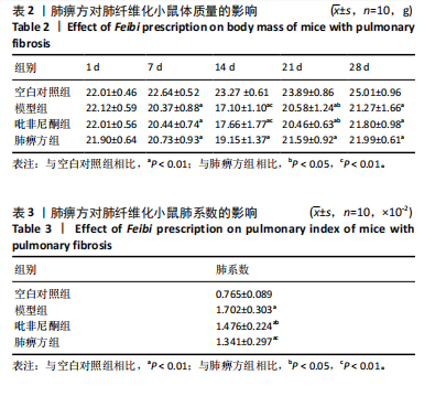
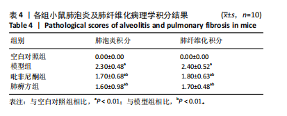
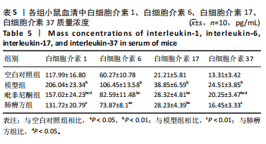
方法:40只C57BL/6雄性小鼠随机分为空白对照组、模型组、吡非尼酮组、肺痹方组,每组10只。除空白对照组外,其他3组腹腔注射博来霉素[7.5 mg/(kg·d)]建立肺纤维化模型,连续注射10 d。造模后第1天各药物组小鼠灌胃给药[51.43/(kg·d)]吡非尼酮或12.86 mg/(kg·d)肺痹方],连续给药28 d。用药结束后取材,采用苏木精-伊红染色和Masson染色观察小鼠肺组织的形态学变化,ELISA法检测血清中白细胞介素1、白细胞介素6、白细胞介素17、白细胞介素37水平,Western-blot法检测肺组织中Bax、Bcl-2、Beclin-1和Caspase3表达。
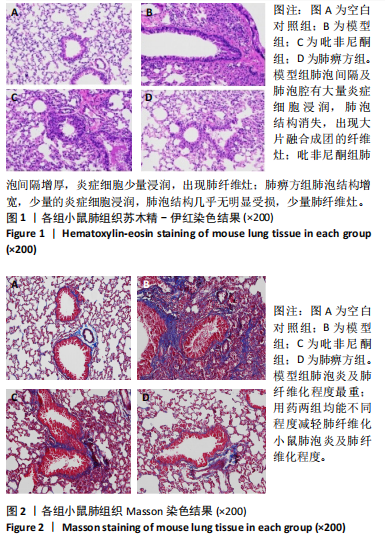
结果与结论:①肺组织的形态学观察显示,模型组肺泡间隔及肺泡腔有大量炎症细胞浸润,出现大片融合成团的纤维灶;吡非尼酮组肺泡间隔增厚,炎症细胞少量浸润,出现肺纤维灶;肺痹方组肺泡结构增宽,少量的炎症细胞浸润,肺泡结构几乎无明显受损,少量肺纤维灶。②与空白对照组相比,模型组小鼠血清白细胞介素1、白细胞介素6、白细胞介素17和白细胞介素37质量浓度明显升高(P < 0.01),两用药组明显低于模型组(P < 0.01),肺痹方组低于非尼酮组。③与空白对照组相比,模型组小鼠肺组织Bax和Caspase3蛋白表达显著升高,两用药组均低于模型组;与空白对照组相比,模型组Bcl-2和Beclin-1蛋白表达显著降低,两用药组均高于模型组。④结论:肺痹方可以减轻肺纤维化,其机制可能与下调白细胞介素1、白细胞介素6、白细胞介素17和白细胞介素37水平,以及调节线粒体凋亡Bax、Bcl-2、Beclin-1和Caspase3相关蛋白从而减少肺泡上皮细胞凋亡有关。
https://orcid.org/0000-0002-7044-5852(程雪)

中国组织工程研究杂志出版内容重点:组织构建;骨细胞;软骨细胞;细胞培养;成纤维细胞;血管内皮细胞;骨质疏松;组织工程

关键词: 肺痹方, 肺纤维化, 线粒体, 博来霉素, C57BL/6 小鼠

Abstract: BACKGROUND: Studies have shown that mitochondrial apoptosis of alveolar epithelial cells plays an important role in the pathogenesis of pulmonary fibrosis, and Feibi prescription can attenuate pulmonary fibrosis and inhibit the transformation of extracellular mechanisms in mice with pulmonary fibrosis.  
OBJECTIVE: To investigate the mechanism of Feibi prescription on mitochondrial apoptpsis of alveolar epithelial cells in bleomycin induced pulmonary fibrosis mice. 
METHODS: Forty male C57BL/6 mice were randomly divided into blank control group, model group, pirfenidone group, and Feibi prescription group. There were 10 mice in each group. Except for the blank control group, the other three groups were intraperitoneally injected with bleomycin (7.5 mg/kg per day) for 10 continuous days to establish the model of pulmonary fibrosis. On day 1 after modeling, the mice in corresponding drug groups were intragastrically administered with pirfenidone (51.43 mg/kg per day) or Feibi prescription (12.86 mg/kg per day). Drug administration lasted for 28 days. Then, morphological changes of lung tissue in mice were observed by hematoxylin-eosin staining and Masson staining. The levels of interleukin-1, interleukin-6, interleukin-17, and interleukin-37 in the serum were detected by ELISA, and the expression of Bax, Bcl-2, Beclin-1, and Caspase3 in the lung tissue was detected by western blot assay. 
RESULTS AND CONCLUSION: Morphological observation of lung tissue showed that in the model group, the alveolar septum and alveolar lumen were infiltrated with a large number of inflammatory cells, and there were large clusters of fibrous foci; in the pirfenidone group, alveolar septa were thickened, with a small infiltration of inflammatory cells and the appearance of pulmonary fibrous foci; in the Feibi prescription group, the alveolar structure was widened, with a small amount of inflammatory cell infiltration, and the alveolar structure was almost not obviously damaged, with a small number of lung fibrous foci. Compared with the blank control group, the mass concentrations of interleukin-1, interleukin-6, interleukin-17, and interleukin-37 were significantly higher in the model group (P < 0.01), while the levels were significantly lower in the two drug groups than the model group (P < 0.01). Moreover, the mass concentrations of interleukin-1, interleukin-6, interleukin-17, and interleukin-37 in the Feibi prescription group were lower than those in the pirfenidone group. Compared with the blank control group, the expression of Bax and Caspase3 proteins in the lung tissue of mice was significantly higher in the model group, while the expression of Bax and Caspase3 proteins was significantly lower in the two drug groups than the model group. Compared with the blank control group, the expression of Bcl-2 and Beclin-1 proteins in the lung tissue of mice was significantly lower in the model group, while the expression of Bcl-2 and Beclin-1 proteins was significantly higher in the two drug groups than the model group. To conclude, Feibi prescription can reduce pulmonary fibrosis and its mechanism may be related to the downregulation of interleukin-1, interleukin-6, interleukin-17, and interleukin-37 levels. This prescription can also reduce the apoptosis of alveolar epithelial cells by regulating mitochondrial apoptosis-related proteins, Bax, Bcl-2, Beclin-1 and Caspase3.

中国组织工程研究杂志出版内容重点:组织构建;骨细胞;软骨细胞;细胞培养;成纤维细胞;血管内皮细胞;骨质疏松;组织工程

Key words: Feibi prescription, pulmonary fibrosis, mitochondrion, bleomycin, C57BL/6 mouse

中图分类号: