[1] LA WG, PARK S, YOON HH, et al. Delivery of a therapeutic protein for bone regeneration from a substrate coated with graphene oxide. Small. 2013;9(23):4051-4060.
[2] DREYER DR, PARK S, BIELAWSKI CW, et al. The chemistry of graphene oxide. Chem Soc Rev. 2010;39(1):228-240.
[3] YANG K, WANG J, CHEN B. Facile fabrication of stable monolayer and few-layer graphene nanosheets as superior sorbents for persistent aromatic pollutant management in water. J Mater Chem A. 2014;2: 18219-18224.
[4] PARK S, AN J, JUNG I, et al. Colloidal suspensions of highly reduced graphene oxide in a wide variety of organic solvents. Nano Lett. 2009; 9(4):1593-1597.
[5] WITHERS N. Graphene oxide: Surfactant sheets. Nat Chem. 2010.https://doi.org/10.1038/nchem.741
[6] ROMERO UAM, SOTO MÁV, JIMÉNEZ LL, et al. Graphene Derivatives: Controlled Properties, Nanocomposites, and Energy Harvesting Applications.G raphene Materials - Structure, Properties and Modifications, 2017.
[7] BÁEZ DF, PARDO H, LABORDA I, et al. Reduced Graphene Oxides: Influence of the Reduction Method on the Electrocatalytic Effect towards Nucleic Acid Oxidation. Nanomaterials (Basel). 2017;7(7):168.
[8] LU C, LU Z, LI Z, et al. Effect of graphene oxide on the mechanical behavior of strain hardening cementitious composites. Constr Build Mater. 2016; 120(1):457-464.
[9] ZHENG Q, HAN B, CUI X, et al. Graphene-Engineered Cementitious Composites: Small Makes a Big Impact. Nanomaterials and Nanotechnology. 2017;7:1-18.
[10] MUNUERA JM, PAREDES JI, VILLAR-RODIL S, et al. High quality, low oxygen content and biocompatible graphene nanosheets obtained by anodic exfoliation of different graphite types. Carbon. 2015;94:729-739.
[11] ZHANG X, YIN J, CHENG P, et al. Distribution and biocompatibility studies of graphene oxide in mice after intravenous administration. Carbon. 2011;49(3):986-995.
[12] YANG K, GONG H, SHI X, et al. In vivo biodistribution and toxicology of functionalized nano-graphene oxide in mice after oral and intraperitoneal administration. Biomaterials. 2013;34(11):2787-2795.
[13] SYAMA S, PAUL W, SABAREESWARAN A, et al. Raman spectroscopy for the detection of organ distribution and clearance of PEGylated reduced graphene oxide and biological consequences. Biomaterials. 2017;131:121-130.
[14] WEI C, LIU Z, JIANG F, et al. Cellular behaviours of bone marrow-derived mesenchymal stem cells towards pristine graphene oxide nanosheets. Cell Prolif. 2017;50(5):e12367.
[15] 付翔,李明新,彭驰伟,等.还原氧化石墨烯对人原代成纤维细胞的毒性研究[J].化学与生物工程,2021,38(2):49-52.
[16] VOLKOV Y, MCINTYRE J, PRINA-MELLO A. Graphene toxicity as a double-edged sword of risks and exploitable opportunities: a critical analysis of the most recent trends and developments. 2D Materials. 2017;4(2):22001.
[17] LI R, GUINEY LM, CHANG CH, et al. Surface Oxidation of Graphene Oxide Determines Membrane Damage, Lipid Peroxidation, and Cytotoxicity in Macrophages in a Pulmonary Toxicity Model. ACS Nano. 2018;12(2):1390-1402.
[18] CAI X, TAN S, LIN M, et al. Synergistic antibacterial brilliant blue/reduced graphene oxide/quaternary phosphonium salt composite with excellent water solubility and specific targeting capability. Langmuir. 2011;27(12):7828-7835.
[19] ZOU X, ZHANG L, WANG Z, et al. Mechanisms of the Antimicrobial Activities of Graphene Materials. J Am Chem Soc. 2016;138(7):2064-2077.
[20] 吴雨宸,刘凯,苏俭生.添加氧化石墨烯对义齿基托树脂细胞毒性及抗菌性能影响的研究[J].口腔颌面修复学杂志,2020,21(3):129-135.
[21] 吴志富,王佐林.氧化石墨烯对牙周致病菌影响的实验研究[J].口腔颌面外科杂志,2019,29(1):23-29.
[22] HE J, ZHU X, QI Z, et al. Killing dental pathogens using antibacterial graphene oxide. ACS Appl Mater Interfaces. 2015;7(9):5605-5611.
[23] VECITIS CD, ZODROW KR, KANG S, et al. Electronic-structure-dependent bacterial cytotoxicity of single-walled carbon nanotubes. ACS Nano. 2010;4(9):5471-5479.
[24] LU X, FENG X, WERBER JR, et al. Enhanced antibacterial activity through the controlled alignment of graphene oxide nanosheets. Proc Natl Acad Sci U S A. 2017;114(46):E9793-E9801.
[25] LIU S, ZENG TH, HOFMANN M, et al. Antibacterial activity of graphite, graphite oxide, graphene oxide, and reduced graphene oxide: membrane and oxidative stress. ACS Nano. 2011;5(9):6971-6980.
[26] DALLAVALLE M, CALVARESI M, BOTTONI A, et al. Graphene can wreak havoc with cell membranes. ACS Appl Mater Interfaces. 2015;7(7): 4406-4414.
[27] WEST JD, MARNETT LJ. Endogenous reactive intermediates as modulators of cell signaling and cell death. Chem Res Toxicol. 2006; 19(2):173-194.
[28] LI J, WANG G, ZHU H, et al. Antibacterial activity of large-area monolayer graphene film manipulated by charge transfer. Sci Rep. 2014;4:4359.
[29] Tu Y, Lv M, Xiu P, et al. Destructive extraction of phospholipids from Escherichia coli membranes by graphene nanosheets. Nat Nanotechnol. 2013;8(8):594-601.
[30] LIU S, HU M, ZENG TH, et al. Lateral dimension-dependent antibacterial activity of graphene oxide sheets. Langmuir. 2012;28(33):12364-12372.
[31] WANG J, WEI Y, SHI X, et al. Cellular entry of graphene nanosheets: the role of thickness, oxidation and surface adsorption. Rsc Advances. 2013;3(36):15776-15782.
[32] MANGADLAO JD, SANTOS CM, FELIPE MJ, et al. On the antibacterial mechanism of graphene oxide (GO) Langmuir-Blodgett films. Chem Commun (Camb). 2015;51(14):2886-2889.
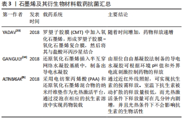
[33] YADAV I, NAYAK SK, RATHNAM VSS, et al. Reinforcing effect of graphene oxide reinforcement on the properties of poly (vinyl alcohol) and carboxymethyl tamarind gum based phase-separated film. J Mech Behav Biomed Mater. 2018;81:61-71.
[34] GANGULY S, DAS P, MAITY PP, et al. Green Reduced Graphene Oxide Toughened Semi-IPN Monolith Hydrogel as Dual Responsive Drug Release System: Rheological, Physicomechanical, and Electrical Evaluations. J Phys Chem B. 2018;122(29):7201-7218.
[35] ALTINBASAK I, JIJIE R, BARRAS A, et al. Reduced Graphene-Oxide-Embedded Polymeric Nanofiber Mats: An “On-Demand” Photothermally Triggered Antibiotic Release Platform. ACS Appl Mater Interfaces. 2018;10(48):41098-41106.
[36] 崔夏青. 氧化石墨烯与石墨烯对成骨细胞增殖分化影响的研究[D].唐山:华北理工大学,2021.
[37] 李婷婷,张玉峰,王若茜,等.石墨烯及其衍生物改性复合材料促成骨机制和应用的研究进展[J].国际口腔医学杂志,2018,45(6):673-677.
[38] 王楠,周延民.石墨烯材料在口腔医学领域的应用及生物安全性研究[J].口腔医学研究,2020,36(5):410-412.
[39] 张敏,周艳,宦泓,等.引导性组织再生联合植骨术对Ⅰ型牙周-牙髓联合病变的临床疗效分析[J].牙体牙髓牙周病学杂志,2018, 28(12):720-723.
[40] 许凌.牙周引导组织再生技术治疗牙周病的临床疗效及影响因素分析[J].罕少疾病杂志,2021,28(1):12-14.
[41] 张岚,吴燕岷.可吸收生物膜在牙周引导组织再生术中的研究进展[J].口腔医学,2020,40(6):571-575.
[42] WANG J, WANG L, ZHOU Z, et al. Biodegradable Polymer Membranes Applied in Guided Bone/Tissue Regeneration: A Review. Polymers (Basel). 2016;8(4):115.
[43] CARBONELL JM, MARTÍN IS, SANTOS A, et al. High-density polytetrafluoroethylene membranes in guided bone and tissue regeneration procedures: a literature review. Int J Oral Maxillofac Surg. 2014;43(1):75-84.
[44] DE MARCO P, ZARA S, DE COLLI M, et al. Graphene oxide improves the biocompatibility of collagen membranes in an in vitro model of human primary gingival fibroblasts. Biomed Mater. 2017;12(5):055005.
[45] 毕博,臧圣奇,何懋典,等.还原氧化石墨烯修饰壳聚糖骨组织工程支架的制备及表征[J].医学研究生学报,2021,34(4):350-356.
[46] 赵彬,武峰,白莹莹,等.负载辛伐他汀的氧化石墨烯/丝素蛋白屏障膜的制备及其生物学性能[J].新型炭材料,2018,33(5):460-468.
[47] 李鹏,木合塔尔·霍加.牙髓干细胞在再生医学中的应用进展[J].全科口腔医学电子杂志,2018,5(24):24,26.
[48] 陈宁馨,赖光云,汪俊.脱落乳牙牙髓干细胞在口腔颌面部组织再生及免疫调节中应用的研究进展[J].中国实用口腔科杂志,2021, 14(2):220-224.
[49] 吴博昊,安莹.牙周膜干细胞在牙周组织再生中的研究新进展[J].口腔生物医学,2020,11(4):270-276.
[50] CHEN FM, SUN HH, LU H, et al. Stem cell-delivery therapeutics for periodontal tissue regeneration. Biomaterials. 2012;33(27):6320-6344.
[51] SEO BM, MIURA M, GRONTHOS S, et al. Investigation of multipotent postnatal stem cells from human periodontal ligament. Lancet. 2004; 364(9429):149-155.
[52] XIE H, CAO T, GOMES JV, et al. Two and three-dimensional graphene substrates to magnify osteogenic differentiation of periodontal ligament stem cells. Carbon. 2015;93:266-275.
[53] ZHOU Q, YANG P, LI X, et al. Bioactivity of periodontal ligament stem cells on sodium titanate coated with graphene oxide. Sci Rep. 2016;6: 19343.
[54] PARK J, PARK S, KIM JE, et al. Enhanced Osteogenic Differentiation of Periodontal Ligament Stem Cells Using a Graphene Oxide-Coated Poly(ε-caprolactone) Scaffold. Polymers (Basel). 2021;13(5):797.
[55] VERA-SÁNCHEZ M, AZNAR-CERVANTES S, JOVER E, et al. Silk-Fibroin and Graphene Oxide Composites Promote Human Periodontal Ligament Stem Cell Spontaneous Differentiation into Osteo/Cementoblast-Like Cells. Stem Cells Dev. 2016;25(22):1742-1754.
[56] TORII D, TSUTSUI TW, WATANABE N, et al. Bone morphogenetic protein 7 induces cementogenic differentiation of human periodontal ligament-derived mesenchymal stem cells. Odontology. 2016;104(1):1-9.
[57] GRONTHOS S, MANKANI M, BRAHIM J, et al. Postnatal human dental pulp stem cells (DPSCs) in vitro and in vivo. Proc Natl Acad Sci U S A. 2000;97(25):13625-13630.
[58] JIA L, GU W, ZHANG Y, et al. The Crosstalk between HDPSCs and HUCMSCs on Proliferation and Osteogenic Genes Expression in Coculture System. Int J Med Sci. 2017;14(11):1118-1129.
[59] XIE H, CHUA M, ISLAM I, et al. CVD-grown monolayer graphene induces osteogenic but not odontoblastic differentiation of dental pulp stem cells. Dent Mater. 2017;33(1):e13-e21.
[60] LEE C, WEI X, KYSAR JW, et al. Measurement of the elastic properties and intrinsic strength of monolayer graphene. Science. 2008; 321(5887):385-388.
[61] LU Q, PANDYA M, RUFAIHAH AJ, et al. Modulation of Dental Pulp Stem Cell Odontogenesis in a Tunable PEG-Fibrinogen Hydrogel System. Stem Cells Int. 2015;2015:525367.
[62] 魏常博,余智超,李晔,等.氧化石墨烯片对人乳牙牙髓干细胞黏附,增殖及成骨早期相关蛋白表达的影响[J]. 临床口腔医学杂志, 2018,34(7):387-391.
[63] 王熙,张淑悦,黄呈森,等.石墨烯对人牙槽骨骨髓间充质干细胞增殖分化的影响[J].卫生职业教育,2020,38(16):117-120.
[64] ZHAO C, LU X, ZANDEN C, et al. The promising application of graphene oxide as coating materials in orthopedic implants: preparation, characterization and cell behavior. Biomed Mater. 2015;10(1):015019.
[65] LEE JH, SHIN YC, JIN OS, et al. Reduced graphene oxide-coated hydroxyapatite composites stimulate spontaneous osteogenic differentiation of human mesenchymal stem cells. Nanoscale. 2015; 7(27):11642-11651.
[66] BREGNOCCHI A, ZANNI E, UCCELLETTI D, et al. Graphene-based dental adhesive with anti-biofilm activity. J Nanobiotechnology. 2017;15(1):89.
[67] 何剑亮.氧化石墨烯对变异链球菌的抗菌作用研究[D].上海:上海交通大学,2016.
|