中国组织工程研究 ›› 2023, Vol. 27 ›› Issue (3): 335-338.doi: 10.12307/2023.021

• 组织工程血管材料 tissue-engineered vascular materials • 上一篇    下一篇

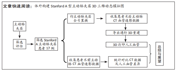
体外构建Stanford A型主动脉夹层3D三维动态模拟图及个体化组织工程血管

程明光1,张超宇2,庄康乐3,阮  鹏1,左  逸1,周正春1,孔  祥1,葛建军1,程光存1   

  1. 1中国科学技术大学附属第一医院(安徽省立医院)心脏外科,安徽省合肥市  230001;2安徽医科大学,安徽省合肥市  230032;3安徽中健三维科技有限公司,安徽省芜湖市  241000
  • 收稿日期:2021-10-12 接受日期:2022-01-13 出版日期:2023-01-28 发布日期:2022-05-20
  • 通讯作者: 程光存,教授,主任医师,中国科学技术大学附属第一医院(安徽省立医院)心脏外科,安徽省合肥市 230001
  • 作者简介:程明光,男,1986年生,安徽省合肥市人,医师,主要从事心脏大血管外科临床与基础工作。
  • 基金资助:
    安徽省重点研究与开发计划(202004j07020018),项目负责人:程光存

In vitro construction of Stanford type A aortic dissection 3D dynamic simulation diagram and individual tissue-engineered blood vessels

Cheng Mingguang1, Zhang Chaoyu2, Zhuang Kangle3, Ruan Peng1, Zuo Yi1, Zhou Zhengchun1, Kong Xiang1, Ge Jianjun1, Cheng Guangcun1   

  1. 1Department of Cardiac Surgery, First Affiliated Hospital (Anhui Provincial Hospital) of University of Science and Technology of China, Hefei 230001, Anhui Province, China; 2Anhui Medical University, Hefei 230032, Anhui Province, China; 3Anhui Zhongjian 3D Technology Co., Ltd., Wuhu 241000, Anhui Province, China
  • Received:2021-10-12 Accepted:2022-01-13 Online:2023-01-28 Published:2022-05-20
  • Contact: Cheng Guangcun, Professor, Chief physician, Department of Cardiac Surgery, First Affiliated Hospital (Anhui Provincial Hospital) of University of Science and Technology of China, Hefei 230001, Anhui Province, China
  • About author:Cheng Mingguang, Physician, Department of Cardiac Surgery, First Affiliated Hospital (Anhui Provincial Hospital) of University of Science and Technology of China, Hefei 230001, Anhui Province, China
  • Supported by:
    Key Research and Development Program of Anhui Province, No. 202004j07020018 (to CGC)

摘要:

文题释义:
个体化组织工程血管:针对个体化患者通过利用生物可降解材料来制备、重建和再生的血管替代材料。
三维动态图:通过计算机辅助设计(CAD)或计算机动画建模软件建模对血管状态进行分析、体外重建。

背景:Stanford A型主动脉夹层手术难度大、复杂程度高,需要在深低温停循环下行升主动脉全弓置换+降主动脉支架置入手术,对术者的操作要求很高。
目的:探讨在体外构建Stanford A型主动脉夹层3D三维动态模拟图及个体化组织工程化血管制备的可行性,为未来为临床医师提供模拟手术过程。
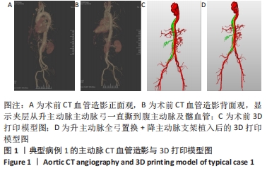
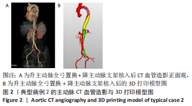
方法:选择2020年1月至2021年7月中国科学技术大学附属第一医院(安徽省立医院)收治的Stanford A型主动脉夹层患者17例,将患者CT影像数据导入到医学图像处理软件,重建出Stanford A型主动脉夹层的主动脉血管及其分支模拟图。对重建出的主动脉血管模型制作出夹层的3D三维动态模拟图,制定个体化组织工程化血管,同时测量主动脉夹层模型与CT血管造影在不同位置的血管内径。
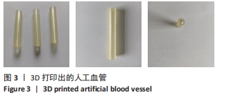
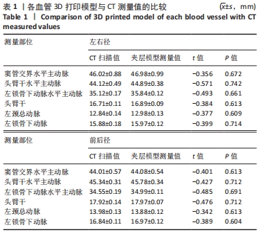
结果与结论:①精确制定出夹层的3D三维动态模拟图和模型,可以制备出个体化的组织工程化血管,满足临床人工血管材料需要;②在窦管交界水平主动脉、头臂干水平主动脉、左锁骨下动脉水平主动脉、头臂干、左颈总动脉、左锁骨下动脉部位,Stanford A型主动脉夹层3D三维动态模型的血管内径值与CT血管造影血管造影检测结果比较差异均无显著性意义(P > 0.05);③结果提示,在体外可构建Stanford A型主动脉夹层3D三维动态模拟图及精确打印出Stanford A型主动脉夹层复杂的解剖结构,可以精确制备个体化组织工程化血管。

https://orcid.org/0000-0002-9347-8091 (程明光)

中国组织工程研究杂志出版内容重点:生物材料;骨生物材料口腔生物材料纳米材料缓释材料材料相容性组织工程

关键词: 主动脉夹层, 3D三维动态模拟图, 夹层模型, 聚乳酸, 组织工程化血管, CT血管造影

Abstract: BACKGROUND: Stanford type A aortic dissection operation is difficult and complex, requiring total ascending aortic arch replacement and descending aortic stent implantation under deep hypothermic circulation arrest. The operator has high operational requirements. 
OBJECTIVE: To explore the feasibility of constructing Stanford type A aortic dissection 3D dynamic simulation diagram in vitro and preparing individual tissue-engineered blood vessels, so as to provide simulated surgical process for clinicians in the future.
METHODS: A total of 17 Stanford type A aortic dissection patients admitted to the First Affiliated Hospital (Anhui Provincial Hospital) of University of Science and Technology of China from January 2020 to July 2021 were selected and their image data were imported into medical image processing software. The simulation of Stanford type A aortic dissection was reconstructed. For the reconstructed aortic vessel model, the 3D dynamic simulation diagram of the dissection was developed, and the individual tissue-engineered vessels were developed. Meanwhile, the average difference between the aortic dissection model and aortic dissection CT angiography at different positions was measured.
RESULTS AND CONCLUSION: (1) Accurate formulation of 3D dynamic simulation diagram and model of interlayer could prepare individual tissue-engineered blood vessels to meet the needs of clinical artificial blood vessel materials. (2) In horizontal aorta at sinus canal junction, horizontal aorta of brachiocephalic trunk, horizontal aorta of left subclavian artery, brachiocephalic trunk, left common carotid artery and left subclavian artery, there was no significant difference between the inner diameter value of the 3D dynamic model of Stanford type A aortic dissection and the results of CT angiography (P > 0.05). (3) It is concluded that constructing 3D dynamic simulation diagram of Stanford type A aortic dissection in vitro and accurately printing the complex anatomical structure of Stanford type A aortic dissection can accurately prepare individual tissue-engineered blood vessels. 

Key words: aortic dissection, 3D dynamic simulation diagram, interlayer model, polylactic acid, tissue-engineered blood vessels, CT angiography

中图分类号: