[1] LU HP, LIU Y, GUO J, et al. Biomaterials with antibacterial and osteoinductive properties to repair infected bone defects. Int J Mol Sci. 2016;17(3):334.
[2] KOSKI C, VU AA, BOSE S. Effects of chitosan-loaded hydroxyapatite on osteoblasts and osteosarcoma for chemopreventative applications. Mater Sci Eng C Mater Biol Appl. 2020;115:111041.
[3] YE YH, PANG YC, ZHANG Z, et al. Decellularized periosteum-covered chitosan globule composite for bone regeneration in rabbit femur condyle bone defects. Macromol Biosci. 2018;18(9):e1700424.
[4] KJALARSDOTTIR L, DYRFJORD A, DAGBJARTSSON A, et al. Bone remodeling effect of a chitosan and calcium phosphate-based composite. Regen Biomater. 2019;6(4):241-247.
[5] TAO FH, MA SJ, TAO H, et al. Chitosan-based drug delivery systems: from synthesis strategy to osteomyelitis treatment - a review. Carbohydr Polym. 2021;251:117063.
[6] ZHAO DY, ZHU TT, LI J, et al. Poly(lactic-co-glycolic acid)-based composite bone-substitute materials. Bioact Mater. 2021;6(2):346-360.
[7] KELLER L, REGIEL-FUTYRA A, GIMENO M, et al. Chitosan-based nanocomposites for the repair of bone defects. Nanomedicine. 2017; 13(7):2231-2240.
[8] RUAN SQ, DENG J, YAN L, et al. Composite scaffolds loaded with bone mesenchymal stem cells promote the repair of radial bone defects in rabbit model. Biomed Pharmacother. 2018; 97:600-606.
[9] ALIDADI S, ORYAN A, BIGHAM-SADEGH A, et al. Comparative study on the healing potential of chitosan, polymethylmethacrylate, and demineralized bone matrix in radial bone defects of rat. Carbohydr Polym. 2017;166:236-248.
[10] ABUEVA CDG, JANG DW, PADALHIN A, et al. Phosphonate-chitosan functionalization of a multi-channel hydroxyapatite scaffold for interfacial implant-bone tissue integration. J Mater Chem B. 2017; 5(6):1293-1301.
[11] ZHANG M, MATINLINNA JP, TSOI JKH, et al. Recent developments in biomaterials for long-bone segmental defect reconstruction: A narrative overview. J Orthop Translat. 2020;22:26-33.
[12] ZHU TT, CUI YT, ZHANG MR, et al. Engineered three-dimensional scaffolds for enhanced bone regeneration in osteonecrosis. Bioact Mater. 2020;5(3):584-601.
[13] AGUILAR A, ZEIN N, HARMOUCH E, et al. Application of chitosan in bone and dental engineering. Molecules. 2019;24(16):3009.
[14] MOSTAFA AA, EL-SAYED MMH, MAHMOUD AA, et al. Bioactive/natural polymeric scaffolds loaded with ciprofloxacin for treatment of osteomyelitis. AAPS PharmSciTech. 2017;18(4):1056-1069.
[15] KYZAS GZ, BIKIARIS DN. Recent modifications of chitosan for adsorption applications: a critical and systematic review. Marine Drugs. 2015; 13(1):312-337.
[16] ZANG SQ, ZHU L, LUO KF, et al. Chitosan composite scaffold combined with bone marrow-derived mesenchymal stem cells for bone regeneration: in vitro and in vivo evaluation. Oncotarget. 2017;8(67): 110890-110903.
[17] TANG YQ, WANG QY, KE QF, et al. Mineralization of ytterbium-doped hydroxyapatite nanorod arrays in magnetic chitosan scaffolds improves osteogenic and angiogenic abilities for bone defect healing. Chem Eng J. 2020;387.https://doi.org/10.1016/j.cej.2020.124166
[18] AAM BB, HEGGSET EB, NORBERG AL, et al. Production of chitooligosaccharides and their potential applications in medicine. Marine Drugs. 2010;8(5):1482-1517.
[19] LI HJ, HU C, YU HJ, et al. Chitosan composite scaffolds for articular cartilage defect repair: a review. RSC Adv. 2018;8(7):3736-3749.
[20] LOGITHKUMAR R, KESHAVNARAYAN A, DHIVYA S, et al. A review of chitosan and its derivatives in bone tissue engineering. Carbohydr Polym. 2016;151:172-188.
[21] CUI LG, ZHANG J, ZOU J, et al. Electroactive composite scaffold with locally expressed osteoinductive factor for synergistic bone repair upon electrical stimulation. Biomaterials. 2020;230:119617.
[22] COVARRUBIAS C, CADIZ M, MAUREIRA M, et al. Bionanocomposite scaffolds based on chitosan-gelatin and nanodimensional bioactive glass particles: in vitro properties and in vivo bone regeneration. J Biomater Appl. 2018;32(9):1155-1163.
[23] MIAO QJ, YANG SY, DING HN, et al. Controlled degradation of chitosan-coated strontium-doped calcium sulfate hemihydrate composite cement promotes bone defect repair in osteoporosis rats. Biomed Mater. 2020;15(5):055039.
[24] MUNHOZ MAS, HIRATA HH, PLEPIS MAG, et al. Use of collagen/chitosan sponges mineralized with hydroxyapatite for the repair of cranial defects in rats. Injury. 2018;49(12):2154-2160.
[25] WANG B, GUO YW, CHEN XF, et al. Nanoparticle-modified chitosan-agarose-gelatin scaffold for sustained release of SDF-1 and BMP-2. Int J Nanomedicine. 2018;13:7395-7408.
[26] JAYASH SN, HASHIM NM, MISRAN M, et al. Formulation and in vitro and in vivo evaluation of a new osteoprotegerin-chitosan gel for bone tissue regeneration. J Biomed Mater Res A. 2017;105(2):398-407.
[27] PETER M, BINULAL NS, NAIR SV, et al. Novel biodegradable chitosan-gelatin/nano-bioactive glass ceramic composite scaffolds for alveolar bone tissue engineering. Chem Eng J. 2010;158(2):353-361.
[28] SARAVANAN S, CHAWLA A, VAIRAMANI M, et al. Scaffolds containing chitosan, gelatin and graphene oxide for bone tissue regeneration in vitro and in vivo. Int J Biol Macromol. 2017;104(Pt B):1975-1985.
[29] YE XL, LI LH, LIN ZF, et al. Integrating 3D-printed PHBV/Calcium sulfate hemihydrate scaffold and chitosan hydrogel for enhanced osteogenic property. Carbohydr Polym. 2018;202:106-114.
[30] LU Y, LI LH, ZHU Y, et al. Multifunctional copper-containing carboxymethyl chitosan/alginate scaffolds for eradicating clinical bacterial infection and promoting bone formation. ACS Appl Mater Interfaces. 2018;10(1):127-138.
[31] ZHAO XJ, ZHOU LY, LI QT, et al. Biomimetic mineralization of carboxymethyl chitosan nanofibers with improved osteogenic activity in vitro and in vivo. Carbohydr Polym. 2018;195:225-234.
[32] TAN HL, AO HY, MA R, et al. In vivo effect of quaternized chitosan-loaded polymethylmethacrylate bone cement on methicillin-resistant staphylococcus epidermidis infection of the tibial metaphysis in a rabbit model. Antimicrob Agents Chemother. 2014;58(10):6016-6023.
[33] 季冬青,孙丹丹,和焕香,等.壳聚糖抗菌活性研究进展[J].辽宁中医药大学学报,2018,20(3):82-85.
[34] RAAFAT D, VON-BARGEN K, HAAS A, et al. Insights into the mode of action of chitosan as an antibacterial compound. Appl Environ Microbiol. 2008;74(12):3764-3773.
[35] CHUNG YC, YEH JY, TSAI CF. Antibacterial characteristics and activity of water-soluble chitosan derivatives prepared by the maillard reaction. Molecules. 2011;16(10):8504-8514.
[36] TAN HL, MA R, LIN CC, et al. Quaternized chitosan as an antimicrobial agent: antimicrobial activity, mechanism of action and biomedical applications in orthopedics. Int J Mol Sci. 2013;14(1):1854-1869.
[37] WANG DY, LIU Y, LIU YL, et al. A dual functional bone-defect-filling material with sequential antibacterial and osteoinductive properties for infected bone defect repair. J Biomed Mater Res A. 2019;107(10):2360-2370.
[38] DAVID N, NALLAIYAN R. Biologically anchored chitosan/gelatin-SrHAP scaffold fabricated on Titanium against chronic osteomyelitis infection. Int J Biol Macromol. 2018;110:206-214.
[39] PATEL KD, EL-FIQI A, LEE HY, et al. Chitosan–nanobioactive glass electrophoretic coatings with bone regenerative and drug delivering potential. J Mater Chem. 2012;22(47). DOI:10.1039/C2JM33830K
[40] FRANK LA, ONZI GR, MORAWSKI AS, et al. Chitosan as a coating material for nanoparticles intended for biomedical applications. React Funct Polym. 2020;147.
[41] NANCY D, RAJENDRAN N. Vancomycin incorporated chitosan/gelatin coatings coupled with TiO2-SrHAP surface modified cp-titanium for osteomyelitis treatment. Int J Biol Macromol. 2018;110:197-205.
[42] CHEN Y, LIU XJ, LIU R, et al. Zero-order controlled release of BMP2-derived peptide P24 from the chitosan scaffold by chemical grafting modification technique for promotion of osteogenesis in vitro and enhancement of bone repair in vivo. Theranostics. 2017;7(5):1072-1087.
[43] MALIK MH, SHAHZADI L, BATOOL R, et al. Thyroxine-loaded chitosan/carboxymethyl cellulose/hydroxyapatite hydrogels enhance angiogenesis in in-ovo experiments. Int J Biol Macromol. 2020;145: 1162-1170.
[44] ZHAO J, SHEN G, LIU CS, et al. Enhanced healing of rat calvarial defects with sulfated chitosan-coated calcium-deficient hydroxyapatite/bone morphogenetic protein 2 scaffolds. Tissue Eng Part A. 2012;18(1-2):
185-197.
[45] YANG Y, CHU LY, YANG SB, et al. Dual-functional 3D-printed composite scaffold for inhibiting bacterial infection and promoting bone regeneration in infected bone defect models. Acta Biomater. 2018;79: 265-275.
[46] LAVANYA K, CHANDRAN SV, BALAGANGADHARAN K, et al. Temperature- and pH-responsive chitosan-based injectable hydrogels for bone tissue engineering. Mater Sci Eng C Mater Biol Appl. 2020;111:110862.
[47] SARAVANAN S, VIMALRAJ S, THANIKAIVELAN P, et al. A review on injectable chitosan/beta glycerophosphate hydrogels for bone tissue regeneration. Int J Biol Macromol. 2019;121:38-54.
[48] YU B, ZHANG YC, LI XM, et al. The use of injectable chitosan/nanohydroxyapatite/collagen composites with bone marrow mesenchymal stem cells to promote ectopic bone formation in vivo. J Nanomater. 2013;2013:1-8.
[49] JAYASH SN, HASHIM NM, MISRAN M, et al. Local application of osteoprotegerin-chitosan gel in critical-sized defects in a rabbit model. PeerJ. 2017;5:e3513.
[50] ZHANG XB, ZHU LX, LV H, et al. Repair of rabbit femoral condyle bone defects with injectable nanohydroxyapatite/chitosan composites. J Mater Sci Mater Med. 2012;23(8):1941-1949.
[51] WANG M, SA Y, LI P, et al. A versatile and injectable poly(methyl methacrylate) cement functionalized with quaternized chitosan-glycerophosphate/nanosized hydroxyapatite hydrogels. Mater Sci Eng C Mater Biol Appl. 2018;90:264-272.
|
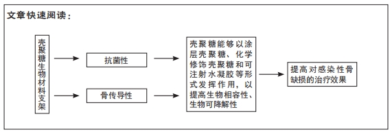

 该文通过总结壳聚糖的骨传导性、抗菌性来回顾最近几年有关壳聚糖生物复合材料支架在治疗感染性骨缺损中的应用研究,同时讨论壳聚糖在骨组织工程中应用的不足之处和其衍生物及复合材料在医学应用中的前景。