[1] DIOMEDE F, MARCONI GD, FONTICOLI L, et al. Functional relationship between osteogenesis and angiogenesis in tissue regeneration. Int J Mol Sci. 2020;21(9): 3242.
[2] HANKENSON KD, DISHOWITZ M, GRAY C, et al. Angiogenesis in bone regeneration. Injury. 2011;42(6):556-561.
[3] KUSUMBE AP, RAMASAMY SK, ITKIN T, et al. Age-dependent modulation of vascular niches for haematopoietic stem cells. Nature. 2016;532(7599):380-384.
[4] DOS SANTOS BP, GARBAY B, FENELON M, et al. Development of a cell-free and growth factor-free hydrogel capable of inducing angiogenesis and innervation after subcutaneous implantation. Acta Biomater. 2019;99:154-167.
[5] LIU Y, OLSEN BR. Distinct vegf functions during bone development and homeostasis. Arch Immunol Ther Exp (Warsz). 2014;62(5):363-368.
[6] BESSA PC, CASAL M, REIS RL. Bone morphogenetic proteins in tissue engineering: The road from laboratory to clinic, part ii (bmp delivery). J Tissue Eng Regen Med. 2008;2(2-3):81-96.
[7] SHARMA S, SAPKOTA D, XUE Y, et al. Delivery of vegfa in bone marrow stromal cells seeded in copolymer scaffold enhances angiogenesis, but is inadequate for osteogenesis as compared with the dual delivery of vegfa and bmp2 in a subcutaneous mouse model. Stem Cell Res Ther. 2018;9(1):23.
[8] SUKUL M, NGUYEN TB, MIN YK, et al. Effect of local sustainable release of bmp2-vegf from nano-cellulose loaded in sponge biphasic calcium phosphate on bone regeneration. Tissue Eng Part A. 2015;21(11-12):1822-1836.
[9] BARATI D, SHARIATI SRP, MOEINZADEH S, et al. Spatiotemporal release of bmp-2 and vegf enhances osteogenic and vasculogenic differentiation of human mesenchymal stem cells and endothelial colony-forming cells co-encapsulated in a patterned hydrogel. J Control Release. 2016;223:126-136.
[10] BAO X, ZHU L, HUANG X, et al. 3d biomimetic artificial bone scaffolds with dual-cytokines spatiotemporal delivery for large weight-bearing bone defect repair. Sci Rep. 2017;7(1):7814.
[11] TANG W, YU Y, WANG J, et al. Enhancement and orchestration of osteogenesis and angiogenesis by a dual-modular design of growth factors delivery scaffolds and 26scs decoration. Biomaterials. 2020;232:119645.
[12] LEE SS, KIM JH, JEONG J, et al. Sequential growth factor releasing double cryogel system for enhanced bone regeneration. Biomaterials. 2020;257:120223.
[13] LARSEN M, WILLEMS WF, PELZER M, et al. Fibroblast growth factor-2 and vascular endothelial growth factor mediated augmentation of angiogenesis and bone formation in vascularized bone allotransplants. Microsurgery. 2014;34(4):301-307.
[14] PATEL JJ, MODES JE, FLANAGAN CL, et al. Dual delivery of epo and bmp2 from a novel modular poly-ɛ-caprolactone construct to increase the bone formation in prefabricated bone flaps. Tissue Eng Part C Methods. 2015;21(9):889-897.
[15] BAYER EA, JORDAN J, ROY A, et al. (*) programmed platelet-derived growth factor-bb and bone morphogenetic protein-2 delivery from a hybrid calcium phosphate/alginate scaffold. Tissue Eng Part A. 2017;23(23-24):1382-1393.
[16] PEREZ RA, KIM JH, BUITRAGO JO, et al. Novel therapeutic core-shell hydrogel scaffolds with sequential delivery of cobalt and bone morphogenetic protein-2 for synergistic bone regeneration. Acta Biomater. 2015;23:295-308.
[17] LI A, LI J, ZHANG Z, et al. Nanohydroxyapatite/polyamide 66 crosslinked with qk and bmp-2-derived peptide prevented femur nonunion in rats. J Mater Chem B. 2021;9(9):2249-2265.
[18] LIM SS, KOOK SH, BHATTARAI G, et al. Local delivery of comp-angiopoietin 1 accelerates new bone formation in rat calvarial defects. J Biomed Mater Res A. 2015;103(9):2942-2951.
[19] PHIPPS MC, XU Y, BELLIS SL. Delivery of platelet-derived growth factor as a chemotactic factor for mesenchymal stem cells by bone-mimetic electrospun scaffolds. PLoS One. 2012;7(7):e40831.
[20] LIU H, LI M, DU L, et al. Local administration of stromal cell-derived factor-1 promotes stem cell recruitment and bone regeneration in a rat periodontal bone defect model. Mater Sci Eng C Mater Biol Appl. 2015;53:83-94.
[21] PAGLIA DN, WEY A, BREITBART EA, et al. Effects of local insulin delivery on subperiosteal angiogenesis and mineralized tissue formation during fracture healing. J Orthop Res. 2013;31(5):783-791.
[22] 冯一梅,徐辉.间充质干细胞基因转染的相关研究现状[J].中国组织工程研究与临床康复,2007,11(11):2118-2121.
[23] ELSHARKASY OM, NORDIN JZ, HAGEY DW, et al. Extracellular vesicles as drug delivery systems: Why and how? Adv Drug Deliv Rev. 2020;159:332-343.
[24] ZHANG Y, HAO Z, WANG P, et al. Exosomes from human umbilical cord mesenchymal stem cells enhance fracture healing through hif-1α-mediated promotion of angiogenesis in a rat model of stabilized fracture. Cell Prolif. 2019; 52(2):e12570.
[25] ZHANG L, JIAO G, REN S, et al. Exosomes from bone marrow mesenchymal stem cells enhance fracture healing through the promotion of osteogenesis and angiogenesis in a rat model of nonunion. Stem Cell Res Ther. 2020;11(1):38.
[26] XIE H, WANG Z, ZHANG L, et al. Extracellular vesicle-functionalized decalcified bone matrix scaffolds with enhanced pro-angiogenic and pro-bone regeneration activities. Sci Rep. 2017;7:45622.
[27] LI H, LIU D, LI C, et al. Exosomes secreted from mutant-hif-1α-modified bone-marrow-derived mesenchymal stem cells attenuate early steroid-induced avascular necrosis of femoral head in rabbit. Cell Biol Int. 2017;41(12):1379-1390.
[28] YING C, WANG R, WANG Z, et al. Bmsc-exosomes carry mutant hif-1α for improving angiogenesis and osteogenesis in critical-sized calvarial defects. Front Bioeng Biotechnol. 2020;8:565561.
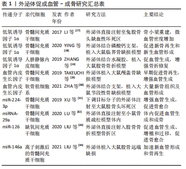
[29] TAKEUCHI R, KATAGIRI W, ENDO S, et al. Exosomes from conditioned media of bone marrow-derived mesenchymal stem cells promote bone regeneration by enhancing angiogenesis. PLoS One. 2019;14(11):e0225472.
[30] ZHA Y, LI Y, LIN T, et al. Progenitor cell-derived exosomes endowed with vegf plasmids enhance osteogenic induction and vascular remodeling in large segmental bone defects. Theranostics. 2021;11(1):397-409.
[31] XU HJ, LIAO W, LIU XZ, et al. Down-regulation of exosomal microrna-224-3p derived from bone marrow-derived mesenchymal stem cells potentiates angiogenesis in traumatic osteonecrosis of the femoral head. FASEB J. 2019;33(7): 8055-8068.
[32] LU GD, CHENG P, LIU T, et al. Bmsc-derived exosomal mir-29a promotes angiogenesis and osteogenesis. Front Cell Dev Biol. 2020;8:608521.
[33] LIU W, LI L, RONG Y, et al. Hypoxic mesenchymal stem cell-derived exosomes promote bone fracture healing by the transfer of mir-126. Acta Biomater. 2020; 103:196-212.
[34] LIU L, YU F, LI L, et al. Bone marrow stromal cells stimulated by strontium-substituted calcium silicate ceramics: Release of exosomal mir-146a regulates osteogenesis and angiogenesis. Acta Biomater. 2021;119:444-457.
[35] BORN LJ, CHANG KH, SHOURESHI P, et al. Hotair-loaded mesenchymal stem/stromal cell extracellular vesicles enhance angiogenesis and wound healing. Adv Healthc Mater. 2021:e2002070.
[36] ZHA Y, LIN T, LI Y, et al. Exosome-mimetics as an engineered gene-activated matrix induces in-situ vascularized osteogenesis. Biomaterials. 2020;247:119985.
[37] SUN P, ZHANG Q, NIE W, et al. Biodegradable mesoporous silica nanocarrier bearing angiogenic qk peptide and dexamethasone for accelerating angiogenesis in bone regeneration. ACS Biomater Sci Eng. 2019;5(12):6766-6778.
[38] BOSE S, FIELDING G, TARAFDER S, et al. Understanding of dopant-induced osteogenesis and angiogenesis in calcium phosphate ceramics. Trends Biotechnol. 2013;31(10):594-605.
[39] ZHENG Z, CHEN Y, HONG H, et al. The “yin and yang” of immunomodulatory magnesium-enriched graphene oxide nanoscrolls decorated biomimetic scaffolds in promoting bone regeneration. Adv Healthc Mater. 2021;10(2):e2000631.
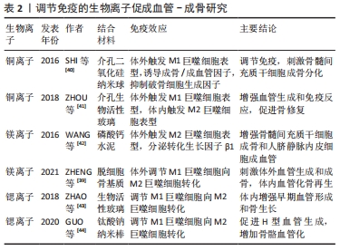
[40] SHI M, CHEN Z, FARNAGHI S, et al. Copper-doped mesoporous silica nanospheres, a promising immunomodulatory agent for inducing osteogenesis. Acta Biomater. 2016;30:334-344.
[41] ZHOU Y, HAN S, XIAO L, et al. Accelerated host angiogenesis and immune responses by ion release from mesoporous bioactive glass. J Mater Chem B. 2018;6(20):3274-3284.
[42] WANG M, YU Y, DAI K, et al. Improved osteogenesis and angiogenesis of magnesium-doped calcium phosphate cement via macrophage immunomodulation. Biomater Sci. 2016;4(11):1574-1583.
[43] ZHAO F, LEI B, LI X, et al. Promoting in vivo early angiogenesis with sub-micrometer strontium-contained bioactive microspheres through modulating macrophage phenotypes. Biomaterials. 2018;178:36-47.
[44] GUO S, YU D, XIAO X, et al. A vessel subtype beneficial for osteogenesis enhanced by strontium-doped sodium titanate nanorods by modulating macrophage polarization. J Mater Chem B. 2020;8(28):6048-6058.
[45] WU C, ZHOU Y, XU M, et al. Copper-containing mesoporous bioactive glass scaffolds with multifunctional properties of angiogenesis capacity, osteostimulation and antibacterial activity. Biomaterials. 2013;34(2):422-433.
[46] ZHANG W, CHANG Q, XU L, et al. Graphene oxide-copper nanocomposite-coated porous cap scaffold for vascularized bone regeneration via activation of hif-1α. Adv Healthc Mater. 2016;5(11):1299-1309.
[47] MAIER JA, BERNARDINI D, RAYSSIGUIER Y, et al. High concentrations of magnesium modulate vascular endothelial cell behaviour in vitro. Biochim Biophys Acta. 2004;1689(1):6-12.
[48] ZHANG Y, XU J, RUAN YC, et al. Implant-derived magnesium induces local neuronal production of cgrp to improve bone-fracture healing in rats. Nat Med. 2016;22(10):1160-1169.
[49] PENG S, LIU X S, HUANG S, et al. The cross-talk between osteoclasts and osteoblasts in response to strontium treatment: Involvement of osteoprotegerin. Bone. 2011;49(6):1290-1298.
[50] CHEN Y, ZHENG Z, ZHOU R, et al. Developing a strontium-releasing graphene oxide-/collagen-based organic-inorganic nanobiocomposite for large bone defect regeneration via mapk signaling pathway. ACS Appl Mater Interfaces. 2019;11(17):15986-15997.
[51] ZHANG W, ZHAO F, HUANG D, et al. Strontium-substituted submicrometer bioactive glasses modulate macrophage responses for improved bone regeneration. ACS Appl Mater Interfaces. 2016;8(45):30747-30758.
[52] D’ANDREA LD, IACCARINO G, FATTORUSSO R, et al. Targeting angiogenesis: Structural characterization and biological properties of a de novo engineered vegf mimicking peptide. Proc Natl Acad Sci U S A. 2005;102(40):14215-14220.
[53] CHEN J, HU G, LI T, et al. Fusion peptide engineered “statically-versatile” titanium implant simultaneously enhancing anti-infection, vascularization and osseointegration. Biomaterials. 2021;264:120446.
[54] SEO E, LIM J S, JUN JB, et al. Exendin-4 in combination with adipose-derived stem cells promotes angiogenesis and improves diabetic wound healing. J Transl Med. 2017;15(1):35.
[55] KANG HM, SOHN I, JUNG J, et al. Exendin-4 protects hindlimb ischemic injury by inducing angiogenesis. Biochem Biophys Res Commun. 2015;465(4):758-763.
[56] QI X, LIU H, MAO L, et al. Combination of exendin-4 and dpp-4 silencing promoted angiogenesis of human coronary artery endothelial cells via activation of pi3k/akt pathway. Pak J Pharm Sci. 2017;30(2(Suppl.)):555-560.
[57] ZHOU H, LI D, SHI C, et al. Effects of exendin-4 on bone marrow mesenchymal stem cell proliferation, migration and apoptosis in vitro. Sci Rep. 2015;5:12898.
[58] MENG J, MA X, WANG N, et al. Activation of glp-1 receptor promotes bone marrow stromal cell osteogenic differentiation through β-catenin. Stem Cell Reports. 2016;6(4):579-591.
[59] ADINI A, ADINI I, CHI ZL, et al. A novel strategy to enhance angiogenesis in vivo using the small vegf-binding peptide pr1p. Angiogenesis. 2017;20(3):399-408.
[60] KIM HK, KIM JH, PARK DS, et al. Osteogenesis induced by a bone forming peptide from the prodomain region of bmp-7. Biomaterials. 2012;33(29):7057-7063.
[61] YU T, LIU Q, JIANG T, et al. Channeled β-tcp scaffolds promoted vascularization and bone augmentation in mandible of beagle dogs. Advanced Functional Materials. 2016;26(37):6719-6727.
[62] WANG H, CHENG H, TANG X, et al. The synergistic effect of bone forming peptide-1 and endothelial progenitor cells to promote vascularization of tissue engineered bone. J Biomed Mater Res A. 2018;106(4):1008-1021.
[63] 周毅,陈跃平,章晓云,等.复方中药圣愈汤治疗股骨头坏死机制的网络药理学分析[J]. 中国组织工程研究,2021,25(17):2687-2696.
[64] GUO Q, YANG J, CHEN Y, et al. Salidroside improves angiogenesis-osteogenesis coupling by regulating the hif-1α/vegf signalling pathway in the bone environment. Eur J Pharmacol. 2020;884:173394.
[65] LIANG S, LING S, DU R, et al. The coupling of reduced type h vessels with unloading-induced bone loss and the protection role of panax quinquefolium saponin in the male mice. Bone. 2021;143:115712.
[66] 黄敏玲,卢赵琦,申震,等.骨碎补总黄酮干预notch信号通路影响骨重建过程中成血管-成骨耦联[J].中国组织工程研究,2021,25(32):5116-5122.
[67] 王晓燕,李冠武,常时新.三七总皂苷通过血管生成改善绝经后骨质疏松机制探析[J].中国骨质疏松杂志,2014,20(8):964-967+977.
[68] 赵志虎,孟新民,孙晓雷,等.柚皮苷对去势大鼠骨折骨痂血管发生的影响及其机制[J].中华骨科杂志, 2016,36(3):177-183.
[69] 武通,陈东宁,许占江.基于vegf/vegfr-2信号通路研究淫羊藿苷促进大鼠胫骨干骨折愈合的作用机制[J].浙江中医药大学学报,2021,45(3):282-288,293.
|