Chinese Journal of Tissue Engineering Research ›› 2013, Vol. 17 ›› Issue (47): 8202-8208.doi: 10.3969/j.issn.2095-4344.2013.47.010
Previous Articles Next Articles
Histocompatibility between small intestinal submucosa and synovial mesenchymal stem cells
Fu Pei-liang, Zhang Lei, Wu Yu-li, Wu Hai-shan, Cong Rui-jun, Chen Song, Ding Zhe-ru, Zhou Qi
- Department of Joint Surgery, Changzheng Hospital of Shanghai, Shanghai 200003, China
-
Revised:2013-08-28Online:2013-11-19Published:2013-11-19 -
Contact:Wu Yu-li, Associate chief physician, Department of Joint Surgery, Changzheng Hospital of Shanghai, Shanghai 200003, China wuyuli6019@189.cn -
About author:Fu Pei-liang☆, M.D., Attending physician, Department of Joint Surgery, Changzheng Hospital of Shanghai, Shanghai 200003, China fupeiliang@163.com Zhang Lei☆, Studying for doctorate, Lecturer, Department of Joint Surgery, Changzheng Hospital of Shanghai, Shanghai 200003, China ra_eagle@hotmail.com Fu Pei-liang and Zhang Lei contributed equally to this work. -
Supported by:the National Natural Science Foundation of China for the Youth, No. 81000798*
CLC Number:
Cite this article
Fu Pei-liang, Zhang Lei, Wu Yu-li, Wu Hai-shan, Cong Rui-jun, Chen Song, Ding Zhe-ru, Zhou Qi. Histocompatibility between small intestinal submucosa and synovial mesenchymal stem cells[J]. Chinese Journal of Tissue Engineering Research, 2013, 17(47): 8202-8208.
share this article
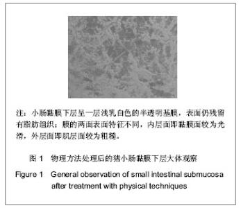
2.1 小肠黏膜下层的制备及检测 大体观察:经物理方法处理后的小肠黏膜下层呈一层浅乳白色的半透明基膜,表面仍残留有脂肪组织;膜的两面表面特征不同,内层面即黏膜面较为光滑,外层面即肌层面较为粗糙,见图1。"

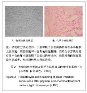
光镜下观察:经物理方法处理过的小肠黏膜下层表面光滑,光学显微镜下细胞计数平均为(30.0±6.1)个/100倍视野,化学方法处理后细胞含量明显下降,平均为(3.0±1.1)个/100倍视野,两者比较差异有显著性意义(P=0.004)。苏木精-伊红染色光镜下观察细胞外基质中有大量胶原纤维,其主要结构和成分没有被破坏,见图2。"
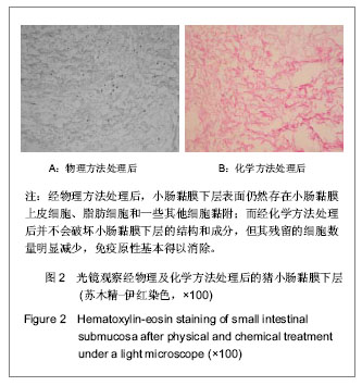
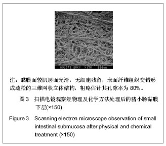
以上结果说明经物理方法处理后,小肠黏膜下层表面仍然存在小肠黏膜上皮细胞、脂肪细胞和一些其他细胞黏附。而经化学方法处理后并不会破坏小肠黏膜下层的结构和成分,但其残留的细胞数量大为减少,免疫原性基本得以消除。扫描电镜下观察:小肠黏膜下层黏膜面较肌层面光滑,无细胞残留,表面纤维组织交错形成疏松的三维网状立体结构,粗略估计其孔隙率为80%,见图3。"
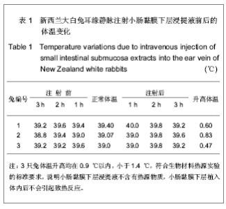
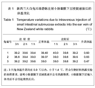
2.2 小肠黏膜下层的生物安全性评价 pH值测定:实验组pH平均值为6.5±0.5,对照组pH值为6.6±0.4,两组间差异无显著性意义(P=0.382)。 热源实验:3只兔体温升高均在0.9 ℃以内,平均(0.6±0.2) ℃,小于1.4 ℃,符合生物材料热源实验的标准要求,说明小肠黏膜下层浸提液不含有热源物质,小肠黏膜下层植入体内后不会引起致热反应,结果见表1。"
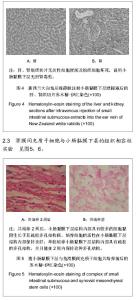
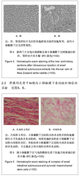
皮肤致敏实验:实验组和阴性对照组在观察期内局部皮肤无红斑、水肿。阳性对照组在24 h时可见红斑、水肿,48 h红斑水肿区扩大,局部皮肤坏死,72 h有焦痂形成。实验证明小肠黏膜下层植入皮内后不会引起致敏反应。 全身毒性实验:实验组和对照组兔在观察期内活动自如,无呼吸急促、呼吸抑制等症状。解剖时未发现腹腔内容物粘连。苏木精-伊红染色肝、肾组织切片无炎性细胞浸润及组织细胞坏死,见图4。"
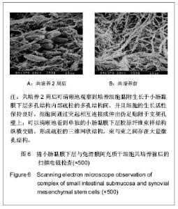
实验组:小肠黏膜下层加入滑膜间充质干细胞再经细胞因子诱导培养2周后的组织切片苏木精-伊红染色光镜观察显示,小肠黏膜下层结构内部具有较多的细胞黏附生长于其疏松多孔结构间,培养细胞的活性在小肠黏膜下层结构内部保持良好,见图5A;扫描电镜可以更进一步清晰地观察到培养细胞黏附生长于小肠黏膜下层多孔结构内部疏松的多孔结构间,并且细胞的生长活性保持良好,细胞间通过突起相互连接或伸出伪足贴附于支架孔壁上,见图6A。 对照组:小肠黏膜下层单独培养2周后的组织切片检查显示,小肠黏膜下层结构内部具有疏松的多孔结构,并且能够在组织培养2周的情况下保持这种多孔结构,见图5B;扫描电镜检查可以清晰地看到胶原纤维束样结构纵横交错,形成疏松三维网状结构,束与束之间存在大量微孔结构,见图6B。"

| [1] 吴海山,徐青镭.膝半月板外科与组织工程学重建[M].上海: 第二军医大学出版社, 1999.
[2] Allman AJ,McPherson TB,Badylak SF,et al.Xenogeneic extracellular matrix grafts elicit a TH2-restricted immune response. Transplantation.2001; 71(11): 1631-1640.
[3] Sarikaya A,Record R,Wu CC,et al.Antimicrobial activity associated with extracellular matrices. Tissue Eng.2002;8(1): 63-71.
[4] Grimes M, Pembroke JT, McGloughlin T. The effect of choice of sterilisation method on the biocompatibility and biodegradability of SIS (small intestinal submucosa). Biomed Mater Eng.2005;15(1-2): 65-71.
[5] Roeder R,Wolfe J,Lianakis N,et al.Compliance, elastic modulus, and burst pressure of small-intestine submucosa (SIS) small-diameter vascular grafts. Boimed Mater Res. 1999;47: 65-70.
[6] Cook JL,Fox DB,Malaviya P,et al.Evaluation of small intestinal submucosa grafts for meniscal regeneration in a clinically relevant posterior meniscectomy model in dogs.J Knee Surg. 2006;19(3):159-167.
[7] 祝云利,荆鑫,缪志和,等.纤维软骨细胞-胶原复合物修复半月板损伤的实验研究[J].解放军医学杂志,2002,27(1): 20-21.
[8] 徐青镭,万年宇,吴海山,等.兔骨髓干细胞向软骨细胞分化用于半月板组织工程重建[J].中国临床康复,2003,7(6): 912-913.
[9] Kobayashi M,Chang YS,Oka M.A two year in vivo study of polyvinyl alcohol-hydrogel (PVA-H) artificial meniscus. Biomaterials.2005;26: 3243-3248.
[10] Bejjani GK,Zabramski J,Durasis Study Group.Safety and efficacy of the porcine small intestinal submucosa dural substitute: results of a prospective multicenter study and literature review. J Neurosurg.2007; 106(6): 1028-1033.
[11] Lawrence BJ,Maase EL,Lin HK,et al.Multilayer composite scaffolds with mechanical properties similar to small intestinal submucosa. J Biomed Mater Res A.2009;88(3): 634-643.
[12] Roeder RA,Lantz GC,Geddes LA.Mechanical remodeling of small-intestine submucosa small-diameter vascular grafts-a preliminary report. Biomed Instrum Technol.2001;35(2): 110-120.
[13] Hadlock TA,Sundback CA,Huntar DA,et al.A new artificial nerve graft containing rolled Schwann cell monolayers. Microsurgery.2001;21(3): 96-101.
[14] Keskin M,Kelly CP,Moreira-Gonzalez A,et al.Repairing critical-sized rat calvarial defects with a periosteal cell-seeded small intestinal submucosal layer. Plast Reconstr Surg.2008; 122(2): 400-409.
[15] Welch JA,Montgomery RD,Lenz SD,et al.Evaluation of small-intestinal submucosa implants for repair of meniscal defects in dogs. Am J Vet Res. 2002;63(3): 427-431.
[16] Prilbitkin EA,Ambro BT,Bloeden E,et al.Rabbit ear cartilage regeneration with a small intestinal submucosa graft. Laryngoscope.2004;114(9 Pt 2 Suppl 102):1-19.
[17] Murphy KD,Mushkudiani IA,Kao D,et al.Successful incorporation of tissue-engineered porcine small-intestinal submucosa as substitute flexor tendon graft is mediated by elevated TGF-beta1 expression in the rabbit. J Hand Surg Am.2008;33(7): 1168-1178.
[18] Derwin K,Androjna C,Spencer E,et al.Porcine small intestine submucosa as a flexor tendon graft. Clin Orthop Relat Res. 2004;423: 245-252.
[19] Musahl V,Abramowitch SD,Gilbert TW,et al.The use of porcine small intestinal submucosa to enhance the healing of the medial collateral ligament--a functional tissue engineering study in rabbits. J Orthop Res. 2004;22(1): 214-220.
[20] Dejardin LM,Arnoczky SP,Ewers BJ,et al.Tissue-engineered rotator cuff tendon using porcine small intestine submucosa: histologic and mechanical evaluation in dogs. Am J Sports Med.200l;29(2):175-184.
[21] Aiken SW,Badylak SF,Toombs JP,et al.Small intestinal submucosa as an intra-articular ligamentous graft material: A pilot study in dogs. Vet Comp Orthoped Traumatol.1994;7: 124-136.
[22] Nihsen ES,Johnson CE,Hiles MC,et a1.Bioactivity of small intestinal submucosa and oxidized regenerated cellulose/collagen. Adv Skin Wound Care. 2008;21(10): 479-486.
[23] Suckow MA,voytik-Harbin SL,Terril LA,et al.Enhanced bone regeneration using porcine small intestinal submucosa. J Invest Surg. 1999;12: 277-287.
[24] Cook JL,Tomlinson JL,Arnoczky SP,et al.Kinetic study of the replacement of porcine small intestinal submucosa grafts and the regeneration of meniscal-like tissue in large avascular meniscal defects in dogs. Tissue Eng.2001;7(3): 321-334.
[25] Rosen M,Ponsky J,Petras R,et al.Small intestinal submucosa as a bioscaffold for biliary tract regeneration. Surgery.2002; 132(3): 480-486.
[26] Record RD,Hillegonds D,Simmons C,et al.In vivo degradation of 14C-labeled small intestinal submucosa(SIS) when used for urinary bladder repair. Biomaterials.2001;22(19): 2653-2659.
[27] Hodde JP,Record RD,Liang HA,et al.Vascular endothelial growth factor in porcine-derived extracellular matrix. Endothelium. 2001;8(1): 11-24.
[28] Voytik-Harbin SL,Brightman AO,Kraine MR,et al.Identification of extractable growth factors from small intestinal submucosa. J Cell Biochem. 1997;67(4): 478-491.
[29] Mcpherson TB,Stephen F.Characterization of fibronectin derived from porcine small intestinal submucosa. Tissue Eng. 1998;4(1): 75-83.
[30] Hodde J,Hiles M. Bioactive FGF-2 in sterilized extracellular matrix. Wounds. 2001;13(5): 195-201.
[31] Abraham GA,Murray J,Billiar K,et al.Evaluation of the porcine intestinal collagen layer as a biomaterial.J Biomed Mater Res. 2000;51(3): 442-452. |
| [1] | He Yan-ping, Ma De-chun, Li Lei, Zhang Li, Zheng Shuang, Dong Ke-xin. Hemocompatibility and surface modification of artificial blood vessel materials [J]. Chinese Journal of Tissue Engineering Research, 2015, 19(8): 1272-1276. |
| [2] | Du Hua, Shi Ying-xu . Bone marrow microenvironment controls the “fate” of hematopoietic stem cells [J]. Chinese Journal of Tissue Engineering Research, 2015, 19(14): 2283-2290. |
| [3] | Wang A-xian, Tang Li, Liang Yuan, Ji Hai-ning, Wu Jun-jie, Ding Yin. Effects of human bone marrow cells-derived extracellular matrix on the proliferation of human periodontal ligament stem cells [J]. Chinese Journal of Tissue Engineering Research, 2014, 18(6): 938-943. |
| [4] | Li Bing-ting, Jia Ying-zhen, Liu Zhi-fang, Song Yuan, Hou Xiao-wei. Effect of freeze-dried bone xenograft and platelet-rich fibrin compound on osteogenesis and osseointegration of alveolar bone defects [J]. Chinese Journal of Tissue Engineering Research, 2014, 18(52): 8376-8381. |
| [5] | Wang Xiao-dong, Zhang Yong-hong. Preparation and performance of recombinant human bone morphogenetic protein-2-poly(hydroxybutyrate-co-hydroxyoctanoate) nanospheres [J]. Chinese Journal of Tissue Engineering Research, 2014, 18(52): 8405-8408. |
| [6] | Feng Chao, Li Zhe, Lv Xiang-guo, Xu Yue-min, Fu Qiang. In vitro preparation and biochemical evaluation of oxygen generative keratin/silk fibroin compound biomaterial [J]. Chinese Journal of Tissue Engineering Research, 2014, 18(52): 8480-8486. |
| [7] | Li Tian-shi, Zeng Wen-ni, He Jun-jun. Chitin and its derivatives inhibit scar formation [J]. Chinese Journal of Tissue Engineering Research, 2014, 18(52): 8504-8508. |
| [8] | Ma Song-feng, Cao Hui, Zheng Feng, Qiao Jun, Zhang Guo-ming. Concomitant cardiac valve replacement and coronary artery bypass grafting [J]. Chinese Journal of Tissue Engineering Research, 2014, 18(5): 699-704. |
| [9] | Liang Yuan, Sui Ke, Shang Feng-qing, Tang Li, Wang A-xian, Ji Hai-ning, Ding Yin. Construction of endothelial progenitor cells/bone marrow mesenchymal stem cells composite sheets [J]. Chinese Journal of Tissue Engineering Research, 2014, 18(41): 6561-6566. |
| [10] | Liu Chen, Wang Sheng-hao, Wang Yi-bin, Tang Xue-bin, Yang Hui-lin, Li Bin. Biocompatibility of annulus fibrosus-derived stem cells with porcine decellularized annulus fibrosus matrix [J]. Chinese Journal of Tissue Engineering Research, 2014, 18(41): 6655-6660. |
| [11] | Lu Hua-ding, Lian Li-yi, Chen Ming-wei, Dai Yu-hu. Cloning and expression of Asperguillus endo-chitosanase gene in Escherichia coli [J]. Chinese Journal of Tissue Engineering Research, 2014, 18(34): 5490-5496. |
| [12] | Fan Zhong-bao, Rong Da-qing, Liu Qing-feng. Prolene hernia system versus polypropylene mesh plug in tension-free hernia repair [J]. Chinese Journal of Tissue Engineering Research, 2014, 18(34): 5535-5539. |
| [13] | Li A-li. Absorbable ligating clip in laparoscopic hysterectomy [J]. Chinese Journal of Tissue Engineering Research, 2014, 18(34): 5561-5565. |
| [14] | Huang Xiao-nan. Percutaneous vertebroplasty for treatment of osteoporotic vertebral fractures: high-viscosity versus low-viscosity bone cement [J]. Chinese Journal of Tissue Engineering Research, 2014, 18(16): 2461-2467. |
| [15] | Liu Li-xia, Chen Lin. Comparison of fluoride release and solubility for different glass ionomer cements [J]. Chinese Journal of Tissue Engineering Research, 2014, 18(16): 2480-2486. |
| Viewed | ||||||
|
Full text |
|
|||||
|
Abstract |
|
|||||