Chinese Journal of Tissue Engineering Research
Previous Articles Next Articles
Application of Accutase enzymes in the separation of spermatogonial stem cells
Liu Shan-shan, Xu Li-ping, Zhu Wei-yun, Ma Ning-fang
- Department of Histology and Embryology, Basic School of Guangzhou Medical University, Guangzhou 510182, Guangdong Province, China
-
Revised:2013-08-19Online:2013-11-05Published:2013-11-05 -
Contact:Ma Ning-fang, M.D., Professor, Department of Histology and Embryology, Basic School of Guangzhou Medical University, Guangzhou 510182, Guangdong Province, China maningfang@yahoo.com.cn -
About author:Liu Shan-shan★, Studying for master’s degree, Department of Histology and Embryology, Basic School of Guangzhou Medical University, Guangzhou 510182, Guangdong Province, China gyliushanshan@163.com -
Supported by:the National Natural Science Foundation of China, No. 81170623*
CLC Number:
Cite this article
Liu Shan-shan, Xu Li-ping, Zhu Wei-yun, Ma Ning-fang. Application of Accutase enzymes in the separation of spermatogonial stem cells[J]. Chinese Journal of Tissue Engineering Research, doi: 10.3969/j.issn.2095-4344.2013.45.013.
share this article

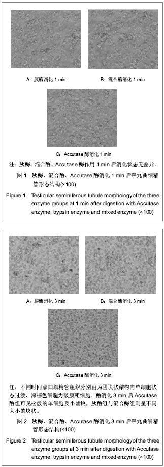
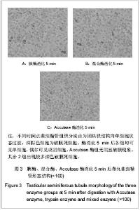
2.1 各组酶的消化作用比较 3组酶消化1,3,5 min后睾丸曲细精管组织结构分别由团块状向单细胞状态过渡,见图1。 各组酶作用1 min后消化状态无明显差异。酶消化 3 min后Accutase酶组明显可见松散的单细胞及小团块,胰酶组与混合酶组则呈不同大小的块状,见图2。"
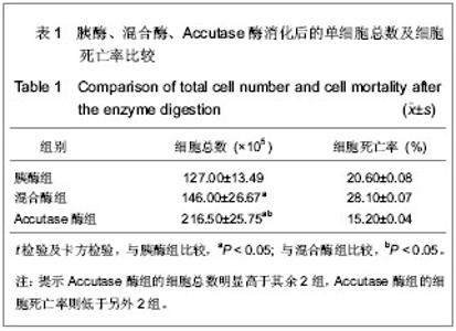
酶消化5 min后各组均可见单细胞,偶尔可见成团细胞,但Accutase酶组细胞完整,无明显破膜现象,而其余2组团块状细胞较多,且出现较多深色破膜死细胞,见图3。"

2.2 单细胞悬液计数及锥虫蓝染色计数 将不同组消化终止后的单细胞悬液分别用细胞计数板计数,并用锥虫蓝染色计数死细胞数,结果见表1。"
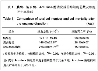
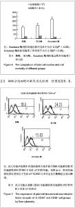
Accutase酶组的细胞总数(216.50±25.75)×105明显高于其余2组(127.00±13.49)×105,(146.00±26.67)× 105;而Accutase酶组的细胞死亡率(15.20±0.04)%则低于另外2组(20.60±0.08)%和(28.10±0.07)%。胰酶组细胞总数低于Accutase酶组,锥虫蓝染色死细胞率高于Accutase酶组(P < 0.05),各组间比较,差异均有显著性意义,见表1,图4。"

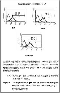
各组酶消化细胞免疫磁珠分选后均可分出CD90+和CD90-两群细胞,流式细胞术分析显示Accutase酶组胶质细胞源性神经营养因子受体α1+/CD90+细胞比率为72.24%,高于胰酶组及混合酶组(51.16%,71.27%);胶质细胞源性神经营养因子受体α1+/CD90-比率为15.03%,明显低于胰酶组及混合酶组(18.8%,24.23%)。"

| [1] Kanatsu-Shinohara M, Inoue K, Lee J, et al. Generation of pluripotent stem cells from neonatal mouse testis. Cell. 2004;119:1001-1012. [2] Guan K, Nayernia K, Maier LS, et al. Pluripotency of spermatogonial stem cells from adult mouse testis. Nature. 2006; 440:1199-1203. [3] Fossa SD, Magelssen H, Melve K, et al. Parenthood in survivors after adulthood cancer and perinatal health in their offspring: a preliminary report. J Natl Cancer Inst Monogr. 2005;34:77-82. [4] Fujita K, Tsujimura A, Miyagawa Y, et al. Isolation of germ cells from leukemia and lymphoma cells in a human in vitro model: potential clinical application for restoring fertility after anticancer therapy. Cancer Res. 2006;66:11166-11171. [5] Seandel M, James D, Shmelkov SV, et al. Generation of functional multipotent adult stem cells from GPR125+ germline progenitors. Nature. 2007;449:346-350. [6] Mitalipova MM, Rao RR, Hoyer DM, et al. Preserving the genetic integrity of human embryonic stem cells. Nat Biotechnol. 2005;23: 19-20. [7] 任萍,关云谦,张愚.人胚皮层神经干细胞培养方法的探讨[J]. 分子细胞生物学报,2007,40(1): 79-83. [8] 黄立宁,韩建民,刘雅.新生大鼠海马神经元原代无血清培养与鉴定[J].基础医学与临床,2012,32(1):83-86. [9] Kanatsu-Shinohara M, Miki H, Inoue K, et al. Long-term culture of mouse male germline stem cells under serum-or feeder-free conditions. Biol Reprod. 2005;72:985-991. [10] Kanatsu-Shinohara M, Takehashi M, Takashima S, et al. Homing of mouse spermatogonial stem cells to germline niche depends on b1-integrin. Cell Stem Cell.2008;3:533-542. [11] 张学明,赖良学,李德雪,等.小鼠精原细胞的分离和纯化[J].解剖学报,2000,31(3):235-238. [12] 李彦锋,郭应禄,李晓红,等.人精原干细胞特异性标志的初步筛选[J].中华男科学杂志,2005,7(11):486-489. [13] Kanatsu-Shinohara M, Lee J, Inoue K, et al. Pluripotency of a single spermatogonial stem cell in mice. Biol Reprod. 2008;78: 681-687. [14] Cyranoski D. Stem cells from testes: could it work? Nature. 2006;440:586-587. [15] Tegelenbosch RA, de Rooij DG. A quantitative study of spermatogonial multiplication and stem cell renewal in the C3H/101 F1 hybrid mouse. Mutat Res. 1993;290:193-200. [16] 张学明,李德雪,李子义,等.小鼠精原干细胞的形态结构特点及其细胞化学和免疫组化特性[J].中国兽医学报, 1999,19(4):363-367. [17] Meng X, Rooij DG, Westerdahl K, et al. Promotion of seminomatous tumors by targeted overexpression of glial cell line-derived neurotrophic factor in mouse testis. Cancer Res. 2001;61(8):3267-3271. [18] Schulz TC, Noggle SA, Palmarini GM, et al. Differentiation of human embryonic stem cells to dopaminergic neurons in serum- free suspension culture. Stem Cells. 2004;22(7):1218-1238. [19] Kallos MS, Behie LA, Vescovi AL. Extended serial passaging of mammalian neural stem cells in suspension bioreactors. Biotechnol Bioeng. 1999;65(5): 589- 599 . [20] Hackett CH, Flaminio MJ, Fortier LA, et al. Analysis of CD14 expression levels in putative mesenchymal progenitor cells isolated from equine bone marrow. Stem Cells Dev. 2011;20: 721-735. [21] Hasegawa K,Fujioka T,Nakamura Y,et al.A method for the selection of human embryonic stemcell sublines with high replating ef?ciency after single-cell dissociation. Stem Cells. 2006;24:2649-2660. [22] Bajpai R, Lesperance J, Kim M, et al. Efficient propagation of single cells Accutase- -dissociated human embryonic stem cells.Mol Reprod Dev. 2008;75(5):818-827. [23] Bajpai R, Lesperance J, Kim M, et al. Efficient propagation of single cells Accutase-dissociated human embryonic stem cells. Mol Reprod Dev. 2008;75(5):818-827. [24] Humason GL. Animal tissue techniques. Freeman San Francisco, 1972: 307-308. [25] Weber C, Pohl S, Pörtner R, et al. Expansion and Harvesting of hMSC-TERT. Open Biomed Eng J. 2007;1:38-146. [26] Kubota H, Avarbock MR, Brinster RL. Culture conditions and single growth factors affect fate determination of mouse spermatogonial stem cells. Biol Reprod. 2004;71:722-731. [27] Kubota H, Avarbock MR, Brinster RL. Growth factors essential for self-renewal and expansion of mouse spermatogonial stem cells. Proc Natl Acad Sci USA. 2004; 101:16489-16494. [28] Kubota H, Avarbock MR, Brinster RL. Spermatogonial stem cells share some, but not all, phenotypic and functional characteristics with other stem cells. Proc Natl Acad Sci U S A. 2003; 100:6487-6492. [29] Meng X, Lindahl M, Hyvönen ME, et al. Regulation of cell fate decision of undifferentiated spermatogonia by GDNF. Science. 2000;287:1489-1493. [30] Jain S, Naughton CK, Yang M, et al. Mice expressing a dominant-negative Ret mutation phenocopy human Hirschsprung disease and delineate a direct role of Ret in spermatogenesis. Development. 2004; 131:5503-5513. [31] Kon J, Ooe H, Oshima H, et al. Expression of CD44 in rat hepatic progenitor cells. J Hepatol. 2006;45: 90-98. [32] Yan AF, Ho DW, Ng MN, et al. Significance of CD90+ cancer stem cells in human liver cancer. Cancer Cell. 2008;13: 153-166. [33] Tiveron C, Marchetti F, Bassani B, et al. Griseofulvin-induced aneuploidy and meiotic delay in female mouse germ cells. I. Cytogenetic analysis of metaphase II oocytes. Mutat Res. 1992; 266(2):143-150. [34] Yao Z,Mishra L. Cancer stem cells and hepatocellular carcinoma. Cancer Biol Ther. 2009;8(18):1691-1698. [35] Tadokoro Y, Yomogida K, Ohta H, et al. Homeostatic regulation of germinal stem cell proliferation by the GDNF/FSH pathway. Mech Develop. 2002;113(1):29-39. [36] Soler RM, Dolcet X, Encinas M, et al. Receptors of the glial cell line-derived neurotrophic factor family of neurotrophic factors signal cell survival through the phosphatidylinositol 3-kinase pathway in spinal cord motoneurons. J Neurosci. 1999;19(21):9160-9169. [37] Naughton CK, Jain S, Strickland AM, et al. Glial cell-line derived neurotrophic factor-mediated RET signaling regulates spermatogonial stem cell fate. Biol Reprod. 2006;74:314-321. [38] Lin LF, Doherty DH, Lile JD, et al. GDNF: a glial cell line-derived neurotrophic factor for midbrain dopaminergic neurons. Science. 1993; 260:1130-1132. [39] Hermann BP, Sukhwani M, Simorangkir DR, et al. Molecular dissection of the male germ cell lineage identifies putative spermatogo-nial stem cells in rhesus macaques. Hum Reprod. 2009;24:1704-1716. [40] Hofmann MC, Braydich-Stolle L, Dym M. Isolation of male germ-line stem cells; influence of GDNF. Dev Biol. 2005;279: 114-124. |
| [1] | Kong Desheng, He Jingjing, Feng Baofeng, Guo Ruiyun, Asiamah Ernest Amponsah, Lü Fei, Zhang Shuhan, Zhang Xiaolin, Ma Jun, Cui Huixian. Efficacy of mesenchymal stem cells in the spinal cord injury of large animal models: a meta-analysis [J]. Chinese Journal of Tissue Engineering Research, 2020, 24(在线): 3-. |
| [2] | Chen Jinsong, Wang Zhonghan, Chang Fei, Liu He. Tissue engineering methods for repair of articular cartilage defect under special conditions [J]. Chinese Journal of Tissue Engineering Research, 2020, 24(8): 1272-1279. |
| [3] | Liu Chundong, Shen Xiaoqing, Zhang Yanli, Zhang Xiaogen, Wu Buling. Effects of strontium-modified titanium surfaces on adhesion, migration and proliferation of bone marrow mesenchymal stem cells and expression of bone formation-related genes [J]. Chinese Journal of Tissue Engineering Research, 2020, 24(7): 1009-1015. |
| [4] | Lin Ming, Pan Jinyong, Zhang Huirong. Knockout of NIPBL gene down-regulates the abilities of proliferation and osteogenic differentiation in mouse bone marrow mesenchymal stem cells [J]. Chinese Journal of Tissue Engineering Research, 2020, 24(7): 1002-1008. |
| [5] | Zhang Wen, Lei Kun, Gao Lei, Li Kuanxin. Neuronal differentiation of rat bone marrow mesenchymal stem cells via lentivirus-mediated bone morphogenetic protein 7 transfection [J]. Chinese Journal of Tissue Engineering Research, 2020, 24(7): 985-990. |
| [6] | Wu Zhifeng, Luo Min. Biomechanical analysis of chemical acellular nerve allograft combined with bone marrow mesenchymal stem cell transplantation for repairing sciatic nerve injury [J]. Chinese Journal of Tissue Engineering Research, 2020, 24(7): 991-995. |
| [7] | Huang Yongming, Huang Qiming, Liu Yanjie, Wang Jun, Cao Zhenwu, Tian Zhenjiang, Chen Bojian, Mai Xiujun, Feng Enhui. Proliferation and apoptosis of chondrocytes co-cultured with TDP43 lentivirus transfected-human umbilical cord mesenchymal stem cells [J]. Chinese Journal of Tissue Engineering Research, 2020, 24(7): 1016-1022. |
| [8] | Qin Xinyu, Zhang Yan, Zhang Ningkun, Gao Lianru, Cheng Tao, Wang Ze, Tong Shanshan, Chen Yu. Elabela promotes differentiation of Wharton’s jelly-derived mesenchymal stem cells into cardiomyocyte-like cells [J]. Chinese Journal of Tissue Engineering Research, 2020, 24(7): 1046-1051. |
| [9] | Liu Mengting, Rao Wei, Han Bing, Xiao Cuihong, Wu Dongcheng. Immunomodulatory characteristics of human umbilical cord mesenchymal stem cells in vitro [J]. Chinese Journal of Tissue Engineering Research, 2020, 24(7): 1063-1068. |
| [10] |
Cen Yanhui, Xia Meng, Jia Wei, Luo Weisheng, Lin Jiang, Chen Songlin, Chen Wei, Liu Peng, Li Mingxing, Li Jingyun, Li Manli, Ai Dingding, Jiang Yunxia.
Baicalein inhibits the biological behavior of hepatocellular
carcinoma stem cells by downregulation of Decoy receptor 3 expression |
| [11] | Zhang Peigen, Heng Xiaolai, Xie Di, Wang Jin, Ma Jinglin, Kang Xuewen. Electrical stimulation combined with neurotrophin 3 promotes proliferation and differentiation of endogenous neural stem cells after spinal cord injury in rats [J]. Chinese Journal of Tissue Engineering Research, 2020, 24(7): 1076-1082. |
| [12] | Huang Cheng, Liu Yuanbing, Dai Yongping, Wang Liangliang, Cui Yihua, Yang Jiandong. Transplantation of bone marrow mesenchymal stem cells overexpressing glial cell line derived neurotrophic factor gene for spinal cord injury [J]. Chinese Journal of Tissue Engineering Research, 2020, 24(7): 1037-1045. |
| [13] | Han Bo, Yang Zhe, Li Jing, Zhang Mingchang . Regulation of limbal stem cells via Wnt signaling in the treatment of limbal stem cell deficiency [J]. Chinese Journal of Tissue Engineering Research, 2020, 24(7): 1057-1062. |
| [14] | Li Jia, Tang Ying, Zhu Qi, Zhang Yanping, Zhou Peigang, Gu Yongchun. Transplantation of human stem cells from the apical papilla for treating dextran sulfate sodium-induced experimental colitis [J]. Chinese Journal of Tissue Engineering Research, 2020, 24(7): 1069-1075. |
| [15] | Zhang Shengmin, Liu Chao. Research progress in osteogenic differentiation of adipose-derived stem cells induced by bioscaffold materials [J]. Chinese Journal of Tissue Engineering Research, 2020, 24(7): 1107-1116. |
| Viewed | ||||||
|
Full text |
|
|||||
|
Abstract |
|
|||||