中国组织工程研究 ›› 2020, Vol. 24 ›› Issue (18): 2873-2878.doi: 10.3969/j.issn.2095-4344.2560

• 骨与关节图像与影像Bone and joint imaging • 上一篇    下一篇

髂骨翼宽度与高度对L5椎弓根置钉遮挡的三维CT评估

张  帅1,欧阳建元1,彭雪莲2,王  松1,王  清1   

  1. 西南医科大学附属医院,1脊柱外科,2超声科,四川省泸州市  646000
  • 收稿日期:2019-09-03 修回日期:2019-09-05 接受日期:2019-10-19 出版日期:2020-06-28 发布日期:2020-04-03
  • 通讯作者: 王清,硕士,主任医师,西南医科大学附属医院脊柱外科,四川省泸州市 646000
  • 作者简介:张帅,男,1992年生,四川省巴中市人,汉族,2018年西南医科大学毕业,硕士,医师,主要从事脊柱外科方面的研究。 共同第一作者:欧阳建元,男,1991年生,四川省绵阳市人,汉族,西南医科大学在读硕士,医师,主要从事脊柱外科方面的研究。

Three-dimensional computed tomography evaluation of L5 pedicle screw fixation shielding by iliac wing width and height 

Zhang Shuai1, Ouyang Jianyuan1, Peng Xuelian2, Wang Song1, Wang Qing1   

  1. 1Department of Spinal Surgery, 2Department of Ultrasound, Affiliated Hospital of Southwest Medical University, Luzhou 646000, Sichuan Province, China
  • Received:2019-09-03 Revised:2019-09-05 Accepted:2019-10-19 Online:2020-06-28 Published:2020-04-03
  • Contact: Wang Qing, Master, Chief physician, Department of Spinal Surgery, Affiliated Hospital of Southwest Medical University, Luzhou 646000, Sichuan Province, China
  • About author:Zhang Shuai, Master, Physician, Department of Spinal Surgery, Affiliated Hospital of Southwest Medical University, Luzhou 646000, Sichuan Province, China Ouyang Jianyuan, Master candidate, Physician, Department of Spinal Surgery, Affiliated Hospital of Southwest Medical University, Luzhou 646000, Sichuan Province, China Zhang Shuai and Ouyang Jianyuan contributed equally to this paper.

摘要:

文题释义:

高髂棘:由于先天发育、创伤、退变等原因导致的双侧髂骨翼达到或超过L4椎弓根下缘称之为高髂棘。

背景:既往学者常根据X射线片对髂棘的高度进行分级。X射线片质量受摄片设备和体位影响大,同时X射线片将髂骨翼与L5椎弓根之间的三维立体关系转变为平面关系,骨性结构重叠使解剖标志辨识困难,尤其老年人常合并骨质疏松、椎旁动脉钙化、肠腔内容物瘀滞等会进一步影响X射线片骨性结构观察。

目的:利用CT三维重建技术观察髂骨翼的宽度和高度对L5椎弓根置钉的遮挡程度。

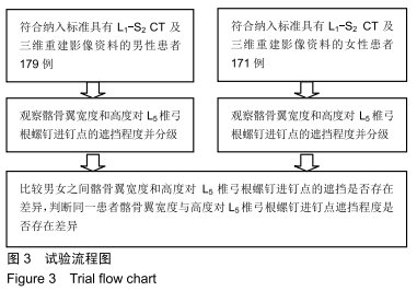
方法:根据纳入和排除标准,选择行L1-S2 CT扫描的350例CT影像资料作为研究对象。所有患者对试验方案均知情同意,且得到医院伦理委员会批准。采用CT三维重建技术在L5椎弓根横轴位中轴层面测量髂骨翼宽度对L5椎弓根螺钉进钉点的遮挡程度,并分为0,Ⅰ,Ⅱ,Ⅲ级;在L5椎弓根斜矢状位中轴层面测量髂骨翼高度对L5椎弓根螺钉进钉点的遮挡程度,同样分为0,Ⅰ,Ⅱ,Ⅲ级。其中0级表示对L5椎弓根螺钉进钉点无遮挡,Ⅰ,Ⅱ,Ⅲ级表示对L5椎弓根螺钉进钉点遮挡程度逐步递增。比较男女之间髂骨翼宽度和高度分别对L5椎弓根螺钉进钉点的遮挡程度是否存在差异。

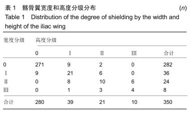
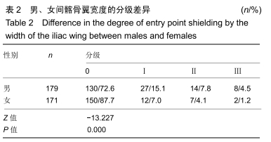
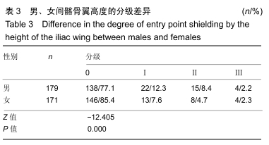
结果与结论:①髂骨翼宽度对L5椎弓根螺钉置钉无遮挡占80.0%(280/350)。阻碍L5椎弓根螺钉置钉占20.0%(70/350),男占27.3%(49/179),其中Ⅰ级27例,Ⅱ级14例,Ⅲ级8例;女占12.3%(21/171),其中Ⅰ级12例,Ⅱ级7例,Ⅲ级2例。②髂骨翼高度对L5椎弓根螺钉置钉无遮挡占80.6%(68/350)。阻碍L5椎弓根螺钉置钉占19.4%(68/350),男占24.0%(43/179),其中Ⅰ级23例,Ⅱ级16例,Ⅲ级4例;女占14.6%(25/171),其中Ⅰ级13例,Ⅱ级8例,Ⅲ级4例。③同一患者髂骨翼宽度对L5椎弓根螺钉横轴位的遮挡和髂骨翼高度对L5椎弓根螺钉矢状位的遮挡程度不完全一致。此组患者中共70例宽髂骨翼,68例高髂骨翼,髂骨翼宽度和高度分级一致共35例,分级不一致达44例;④男性髂骨翼宽度和高度对L5椎弓根螺钉进钉点的遮挡程度均大于女性。⑤结果证实,髂骨翼宽度和高度对L5椎弓根螺钉置钉遮挡发生率分别为20.0%和19.4%。男性髂骨翼宽度和高度对L5椎弓根螺钉进钉点的遮挡程度均大于女性。髂骨翼宽度在横轴位对L5椎弓根螺钉进钉点的遮挡程度与髂骨翼高度在斜矢状位对L5椎弓根螺钉进钉点的遮挡程度并不完全一致,术前采用CT三维重建技术判断髂骨翼与L5椎弓根螺钉进钉点的关系对于提高L5椎弓根置钉安全性及手术决策具有重要意义。

ORCID: 0000-0001-5579-4783(张帅)

中国组织工程研究杂志出版内容重点:人工关节;骨植入物;脊柱骨折;内固定;数字化骨科;组织工程

关键词: 髂骨翼宽度, 髂骨翼高度, L5椎弓根螺钉, CT三维重建, 遮挡

Abstract:

BACKGROUND: Previous scholars often graded the height of iliac spine according to X-ray films. The quality of X-ray images is greatly affected by the photographic equipment and position. Simultaneously, X-ray film transforms the three-dimensional relationship between the iliac wing and the L5 pedicle into a planar relationship. The overlap of bone structure makes it difficult to identify anatomic markers. Especially, the combination with osteoporosis, calcification of paravertebral artery, and stasis of intestinal contents in the elderly will further affect the observation of bone structure on X-ray films.

OBJECTIVE: To observe the degree of the entry point in L5 pedicle screw fixation shielded by the width and height of the iliac wing using the three-dimensional computed tomography reconstruction technique.

METHODS: According to the inclusion and exclusion criteria, 350 CT images of L1-S2 region were selected as the study object. All patients signed the informed consent. This study was approved by the Hospital Ethics Committee. The degree of the entry point in L5 pedicle screw fixation shielded by the width of the iliac wing in central axis layer of the horizontal axis was analyzed using the three-dimensional computed tomography reconstruction technique and divided into levels 0, I, II, and III. The degree of the entry point in L5 pedicle screw fixation shielded by the height of the iliac wing in central axis layer of the oblique sagittal plane was analyzed and also divided into levels 0, I, II, and III. Level 0 indicates that the entry point of L5 pedicle screw is not shielded by the iliac wing, and levels I, II, and III indicate increasing degrees of entry point of L5 pedicle screw shielding by the iliac wing. The difference in the effect of width and height of the iliac wing on the shielding degree of entry point in L5 pedicle screw was compared between males and females. 

RESULTS AND CONCLUSION: (1) In 80.0% of patients (280/350), the entry point of L5 pedicle screw was not shielded by the width of the iliac wing. In 20.0% (70/350) of patients, the entry point of L5 pedicle screw was shielded by the width of the iliac wing. In all, 27.3% (49/179) were male, including 27 with level I shielding, 14 with level II shielding, and 8 with level III shielding; 12.3% (21/171) were female, including 12 with level I shielding, 7 with level II shielding, and 2 with level III shielding. (2) In 80.6% (68/350) of patients, the entry point of L5 pedicle screw was not shielded by the height of the iliac wing. In 19.4% (68/350) of patients, the entry point of L5 pedicle screw was shielded by the height of the iliac wing. In all, 24.0% (43/179) of these patients were male, including 23 cases with level I shielding, 16 cases with level II shielding, and 4 with level III shielding; 14.6% (25/171) were female, including 13 with level I shielding, 8 with level II shielding, and 4 with level III shielding. (3) The degree of entry point of L5 pedicle screw shielding by the width of the iliac wing on the horizontal axis was not exactly the same as that of the height of the iliac wing on the sagittal axis of the L5 pedicle screw in the same patient. In this study, there were 70 patients with wide iliac wings and 68 patients with high iliac wings; the degrees of shielding by the width and height of the iliac wing were the same in 35 cases and different in 44 cases. (4) The degree of entry point of L5 pedicle screw shielding by the width and height of the iliac wing was greater in males than in females. (5) The results confirmed that the incidence of entry point of L5 pedicle screw shielding by the iliac wing width and height was 20.0% and 19.4%, respectively. The degree of entry point of L5 pedicle screw shielding by the width and height of the iliac wing was greater in males than females. The degree of entry point of L5 pedicle screw shielding by the iliac wing width along the horizontal axis was not completely consistent with that of L5 pedicle screw shielding on the oblique sagittal plane. It is of great significance to evaluate the relationship between iliac wing and L5 pedicle screw entry point by three-dimensional computed tomography reconstruction before operation for improving the safety of L5 pedicle screw placement and decision-making of operation.

Key words: iliac wing width, iliac wing height, L5 pedicle screw, three-dimensional computed tomography reconstruction, shielding

中图分类号: